

Arteriovenous malformation presenting as traumatic subdural hematoma: A case report
- PMID: 32874706
- PMCID: PMC7451141
- DOI: 10.25259/SNI_160_2019
Arteriovenous malformation presenting as traumatic subdural hematoma: A case report
Abstract
Background: Brain arteriovenous malformations (AVMs) are congenital aberrant connections between afferent arteries and draining veins with no intervening capillary bed or neural parenchyma. Other than seizures, the most common initial presentation of AVM is hemorrhage, which is typically intraparenchymal, subarachnoid, or intraventricular, and very rarely subdural.
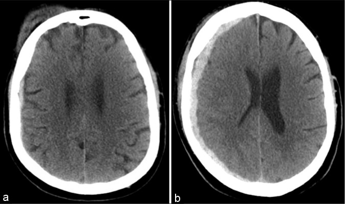
Case description: This patient is a 66-year-old male with a history of atrial fibrillation, chronically anticoagulated with apixaban, who presented through emergency services after a fall. On presentation, computed tomography (CT) of the head showed a small, 6 mm right subdural hematoma, and the patient was neurologically intact. The hematoma was evacuated by burr hole craniotomy and placement of a subdural drain 12 days after the initial presentation due to worsening headaches and further hematoma expansion. Two weeks postevacuation, the patient was readmitted for seizures, and at this time, CT angiography showed no intracranial vascular lesion. Approximately 1 month later, the patient was readmitted for decreased responsiveness, and CT head at this time found right frontal intraparenchymal hemorrhage. On subsequent catheter angiography, the right frontal AVM was discovered. It was treated with preoperative embolization followed by surgical resection. Postoperatively, the patient followed commands and tracked with his eyes. There was spontaneous antigravity movement of the right upper extremity, but still no movement of the left upper or bilateral lower extremities.
Conclusion: This case emphasizes the importance of maintaining a high index of suspicion for underlying vascular lesions when evaluating intracranial bleeding, even in the setting of traumatic history, particularly in cases of hematoma expansion.
Keywords: Arteriovenous malformation; Intracranial hemorrhage; Subdural hematoma.
Copyright: © 2020 Surgical Neurology International.
Conflict of interest statement
There are no conflicts of interest.
Figures


References
-
- Abecassis IJ, Xu DS, Batjer HH, Bendok BR. Natural history of brain arteriovenous malformations: A systematic review. Neurosurg Focus. 2014;37:E7. - PubMed
-
- Al-Shahi R, Bhattacharya JJ, Currie DG, Papanastassiou V, Ritchie V, Roberts RC, et al. Prospective, population-based detection of intracranial vascular malformations in adults: The scottish intracranial vascular malformation study (SIVMS) Stroke. 2003;34:1163–9. - PubMed
-
- Aoun SG, Bendok BR, Batjer HH. Acute management of ruptured arteriovenous malformations and dural arteriovenous fistulas. Neurosurg Clin N Am. 2012;23:87–103. - PubMed
-
- Atkinson RP, Awad IA, Batjer HH, Dowd CF, Furlan A, Giannotta SL, et al. Reporting terminology for brain arteriovenous malformation clinical and radiographic features for use in clinical trials. Stroke. 2001;32:1430–42. - PubMed
-
- Biffl WL, Moore EE, Offner PJ, Brega KE, Franciose RJ, Elliott JP, et al. Optimizing screening for blunt cerebrovascular injuries. Am J Surg. 1999;178:517–22. - PubMed
Publication types
LinkOut - more resources
Full Text Sources