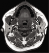
Trigeminal schwannoma presenting with malocclusion: A case report and review of the literature
- PMID: 32874733
- PMCID: PMC7451153
- DOI: 10.25259/SNI_482_2019
Trigeminal schwannoma presenting with malocclusion: A case report and review of the literature
Abstract
Background: Trigeminal schwannomas are rare tumors of the trigeminal nerve. Depending on the location, from which they arise along the trigeminal nerve, these tumors can present with a variety of symptoms that include, but are not limited to, changes in facial sensation, weakness of the masticatory muscles, and facial pain.
Case description: We present a case of a 16-year-old boy with an atypical presentation of a large trigeminal schwannoma: painless malocclusion and unilateral masticatory weakness. This case is the first documented instance; to the best of our knowledge, in which a trigeminal schwannoma has led to underbite malocclusion; it is the 19th documented case of unilateral trigeminal motor neuropathy of any etiology. We discuss this case as a unique presentation of this pathology, and the relevant anatomy implicated in clinical examination aid in further understanding trigeminal nerve pathology.
Conclusion: We believe our patient's underbite malocclusion occurred secondary to his trigeminal schwannoma, resulting in associated atrophy and weakness of the muscles innervated by the mandibular branch of the trigeminal nerve. Furthermore, understanding the trigeminal nerve anatomy is crucial in localizing lesions of the trigeminal nerve.
Keywords: Cranial neuropathy; Malocclusion; Pediatric neurosurgery; Skull base tumor; Trigeminal schwannoma.
Copyright: © 2020 Surgical Neurology International.
Conflict of interest statement
There are no conflicts of interest.
Figures


References
-
- Andonopoulos A, Lagos G, Drosos A, Moutsopoulos H. The spectrum of neurological involvement in Sjögren’s syndrome. Br J Rheumatol. 1990;29:21–4. - PubMed
-
- Bathla G, Hegde A. The trigeminal nerve: An illustrated review of its imaging anatomy and pathology. Clin Radiol. 2013;68:203–13. - PubMed
-
- Beydoun SR. Unilateral trigeminal motor neuropathy as a presenting feature of neurofibromatosis Type 2 (NF2) Muscle Nerve. 1993;16:1136–7. - PubMed
Publication types
LinkOut - more resources
Full Text Sources
