. 2020 Jul 23;9(8):e1171-e1180.
doi: 10.1016/j.eats.2020.04.017. eCollection 2020 Aug.

Arthroscopic "Bone Block Cerclage" Technique for Posterior Shoulder Instability

Affiliations

Arthroscopic "Bone Block Cerclage" Technique for Posterior Shoulder Instability

Abdul-Ilah Hachem et al. Arthrosc Tech. .

Abstract

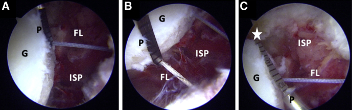
Many open and arthroscopic techniques have been described to treat posterior glenohumeral instability. Multifactorial features of posterior shoulder instability pathoanatomy and varied patient characteristics have challenged the understanding of this condition and have led to dissimilar results, without a strong consensus for the most adequate technique to treat it. We describe an arthroscopic anatomical metal-free posterior glenoid reconstruction technique, using a tricortical iliac crest allograft with 2 ultra-high strength sutures (FiberTape Cerclage System; Arthrex, Naples, FL) with concomitant posterior capsulolabral complex reconstruction procedure.


Figures

Fig 1
Fig 1
Right shoulder, lateral decubitus position. Arthroscopic view, anterosuperior portal. (A) Posteroinferior labral lesion as seen from the anterosuperior portal. (B) Release of the posterior capsulolabral complex and debridement of the posterior glenoid rim. (C, capsule; G, glenoid; HH, humeral head; L, posterior labrum.)
Fig 2
Fig 2
Right shoulder, lateral decubitus position. Arthroscopic view, anterosuperior portal. (A) Posterior capsulolabral complex retraction with PDS. (B) Visualization of the infraspinatus muscle fibers. (C) Lateral decubitus, right shoulder, extra-articular view of the PDS suture fixation. (AP, accessory posterior portal; C, capsule; G, glenoid; HH, humeral head; P, posterior portal; PDS, polydioxanone suture; SSC, subscapularis.)
Fig 3
Fig 3
Right shoulder, lateral decubitus position, Arthroscopic view, anterosuperior portal. Intraoperative measurement of the superoinferior length of the posterior glenoid. White Star: arthroscopic measurement probe, 220 mm, 60°; Arthrex). (C, capsule; G, glenoid; HH, humeral head; ISP, infraspinatus muscle fibers; P, posterior portal; PDS, polydioxanone suture; SSC, subscapularis.)
Fig 4
Fig 4
Right shoulder, lateral decubitus position. Arthroscopic view, anterosuperior portal. (A) Posterior placement of the drilling guide. (B) Lateral decubitus, extra-articular view of the posterior guide insertion. (G, posterior glenoid; P, posterior view of the right shoulder; PG, posterior drilling guide.)
Fig 5
Fig 5
Right shoulder, lateral decubitus position. Arthroscopic view, anterosuperior portal. (A-B) Measure of the distance between articular margin and graft holes. (C) Measure of the distance between inferior graft hole and the distal margin of the glenoid. White star: Distal margin of the glenoid. (FL, FiberLink; G, glenoid; ISP, infraspinatus muscle fibers; P, arthroscopic probe.)
Fig 6
Fig 6
Iliac crest allograft preparation. (A) Allograft measurement and (B) cutting. (C) Allograft orientation. Black star: cortical side of the allograft. (G, glenoid face of the allograft; I, inferior aspect of the allograft; ICA, iliac crest allograft; IT, inferior tunnel; S, superior aspect of the allograft; ST, superior tunnel.)
Fig 7
Fig 7
Right shoulder, lateral decubitus position. Arthroscopic view, anterosuperior portal. (A) Nitinol loop retrieved from the cannulated drill through the anterior portal. (Black arrow pointing to the inferior nitinol loop). (B) Nitinol wires are replaced with 2 different-colored FiberLink sutures. (AL, anterior labrum; C, posterior capsule; FL, FiberLinks; G, posterior glenoid; HH, humeral head; SSC, subscapular.)
Fig 8
Fig 8
Right shoulder, lateral decubitus position. Accommodation of the allograft. (A) Two bands of FiberTape cerclage sutures are passed through the superior allograft hole. (B-C) The FiberTapes are connected with posterior loop of the superior FiberLink and retrieved from the posterior to anterior side of the glenoid. (D-F) The FiberTape sutures are then retrieved from the anterior glenoid hole to the posterior end pulling the inferior FiberLink loop through the glenoid and passing through the inferior drill hole of the graft. (B, D, E) Arthroscopic view from the anterosuperior portal. (F) Intra-articular view. (A, cortical side of the allograft; AC, anterior capsule; FL, FiberLink; FT, FiberTape; G, glenoid; P, posteromedial portal).
Fig 9
Fig 9
Insertion of the allograft into the joint. (A) Lateral decubitus, right shoulder. Extra-articular view of the construct previous insertion. (B) Arthroscopic view from anterosuperior portal. Insertion of allograft through posteromedial portal. (A, allograft; AP, accessory posterior portal; C, posterior capsule; FT, FiberTapes; G, glenoid; P, posterior portal.)
Fig 10
Fig 10
Right shoulder, lateral decubitus position. (A-D) Extra-articular view of the FiberTape interconnection. (AP, accessory posterior portal; FTC, FiberTape cerclage; TTC, TigerTape cerclage.)
Fig 11
Fig 11
Fixation of the allograft bone block, right shoulder, lateral decubitus position. (A) Arthroscopic view from the anterosuperior portal and (B) extra-articular posterior view of the fixation of the allograft with the tensioner. (C) Arthroscopic view from the anterosuperior portal of the bone block fixation after knot tying. (A, allograft; CLC, capsulolabral complex; G, glenoid; K, knots; T, tensioner.)
Fig 12
Fig 12
(A-B) Right shoulder, lateral decubitus position, arthroscopic view from the anterosuperior portal. Capsulolabral complex reconstruction to its native glenoid with FiberTak knotless 1.8 implants (Arthrex) (A, allograft; CLC, capsulolabral complex, G, glenoid; HH, humeral head; I, implant from the posterior percutaneous polydioxanone suture traction portal.)
Fig 13
Fig 13
(A-B) Graphical representation of the posterior bone block cerclage. (A, allograft; FT, FiberTape Cerclage System; G, glenoid.)

References

    1. Woodmass J.M., Lee J., Wu I.T. Incidence of posterior shoulder instability and trends in surgical reconstruction: A 22-year population-based study. J Shoulder Elbow Surg. 2019;28:611–616. - PubMed
    1. Provencher M.T., Leclere L.E., King S. Posterior instability of the shoulder: Diagnosis and management. Am J Sports Med. 2011;39:874–886. - PubMed
    1. Chang E.S., Greco N.J., McClincy M.P., Bradley J.P. Posterior shoulder instability in overhead athletes. Orthop Clin North Am. 2016;47:179–187. - PubMed
    1. Rouleau D.M., Hebert-Davies J., Robinson C.M. Acute traumatic posterior shoulder dislocation. J Am Acad Orthop Surg. 2014;22:145–152. - PubMed
    1. Frank R.M., Romeo A.A., Provencher M.T. Posterior glenohumeral instability: Evidence-based treatment. J Am Acad Orthop Surg. 2017;25:610–623. - PubMed