

Tough and tunable scaffold-hydrogel composite biomaterial for soft-to-hard musculoskeletal tissue interfaces
- PMID: 32875114
- PMCID: PMC7438087
- DOI: 10.1126/sciadv.abb6763
Tough and tunable scaffold-hydrogel composite biomaterial for soft-to-hard musculoskeletal tissue interfaces
Abstract
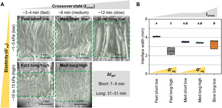
Tendon inserts into bone via a fibrocartilaginous interface (enthesis) that reduces mechanical strain and tissue failure. Despite this toughening mechanism, tears occur because of acute (overload) or degradative (aging) processes. Surgically fixating torn tendon into bone results in the formation of a scar tissue interface with inferior biomechanical properties. Progress toward enthesis regeneration requires biomaterial approaches to protect cells from high levels of interfacial strain. We report an innovative tissue reinforcement strategy: a stratified scaffold containing osseous and tendinous tissue compartments attached through a continuous polyethylene glycol (PEG) hydrogel interface. Tuning the gelation kinetics of the hydrogel modulates integration with the flanking compartments and yields biomechanical performance advantages. Notably, the hydrogel interface reduces formation of strain concentrations between tissue compartments in conventional stratified biomaterials that can have deleterious biological effects. This design of mechanically robust stratified composite biomaterials may be appropriate for a broad range of tendon and ligament-to-bone insertions.
Copyright © 2020 The Authors, some rights reserved; exclusive licensee American Association for the Advancement of Science. No claim to original U.S. Government Works. Distributed under a Creative Commons Attribution NonCommercial License 4.0 (CC BY-NC).
Figures


Similar articles
-
Shaping the mechanical properties of a gelatin hydrogel interface via amination.bioRxiv [Preprint]. 2024 Oct 15:2024.10.14.618299. doi: 10.1101/2024.10.14.618299. bioRxiv. 2024. PMID: 39464090 Free PMC article. Preprint.
-
Effect of kartogenin-loaded gelatin methacryloyl hydrogel scaffold with bone marrow stimulation for enthesis healing in rotator cuff repair.J Shoulder Elbow Surg. 2021 Mar;30(3):544-553. doi: 10.1016/j.jse.2020.06.013. Epub 2020 Jul 7. J Shoulder Elbow Surg. 2021. PMID: 32650072
-
Integrating soft and hard tissues via interface tissue engineering.J Orthop Res. 2018 Apr;36(4):1069-1077. doi: 10.1002/jor.23810. Epub 2018 Jan 5. J Orthop Res. 2018. PMID: 29149506 Free PMC article. Review.
-
Bone-Adhesive Anisotropic Tough Hydrogel Mimicking Tendon Enthesis.Adv Mater. 2023 Jan;35(3):e2206207. doi: 10.1002/adma.202206207. Epub 2022 Dec 14. Adv Mater. 2023. PMID: 36314423
-
Scaffold-based tissue engineering strategies for soft-hard interface regeneration.Regen Biomater. 2022 Nov 12;10:rbac091. doi: 10.1093/rb/rbac091. eCollection 2023. Regen Biomater. 2022. PMID: 36683751 Free PMC article. Review.
Cited by
-
Biomimetic Approaches for the Design and Fabrication of Bone-to-Soft Tissue Interfaces.ACS Biomater Sci Eng. 2023 Jul 10;9(7):3810-3831. doi: 10.1021/acsbiomaterials.1c00620. Epub 2021 Nov 16. ACS Biomater Sci Eng. 2023. PMID: 34784181 Free PMC article. Review.
-
Dynamic Chemistry Toolbox for Advanced Sustainable Materials.Adv Sci (Weinh). 2024 Apr;11(14):e2308666. doi: 10.1002/advs.202308666. Epub 2024 Feb 6. Adv Sci (Weinh). 2024. PMID: 38321810 Free PMC article. Review.
-
Linguistic Analysis Identifies Emergent Biomaterial Fabrication Trends for Orthopaedic Applications.Adv Healthc Mater. 2023 Apr;12(10):e2202591. doi: 10.1002/adhm.202202591. Epub 2023 Feb 5. Adv Healthc Mater. 2023. PMID: 36657736 Free PMC article. Review.
-
Growing Pains: The Need for Engineered Platforms to Study Growth Plate Biology.Adv Healthc Mater. 2022 Oct;11(19):e2200471. doi: 10.1002/adhm.202200471. Epub 2022 Aug 15. Adv Healthc Mater. 2022. PMID: 35905390 Free PMC article. Review.
-
Engineering an enthesis-like graft for rotator cuff repair: An approach to fabricate highly biomimetic scaffold capable of zone-specifically releasing stem cell differentiation inducers.Bioact Mater. 2022 Jan 5;16:451-471. doi: 10.1016/j.bioactmat.2021.12.021. eCollection 2022 Oct. Bioact Mater. 2022. PMID: 35386315 Free PMC article.
References
Publication types
Grants and funding
LinkOut - more resources
Full Text Sources
