Review
. 2020 Aug 24;5(8):857-869.
doi: 10.1016/j.jacbts.2020.04.014. eCollection 2020 Aug.

Autosis: A New Target to Prevent Cell Death

Affiliations
Review

Autosis: A New Target to Prevent Cell Death

Jihoon Nah et al. JACC Basic Transl Sci. .

Abstract

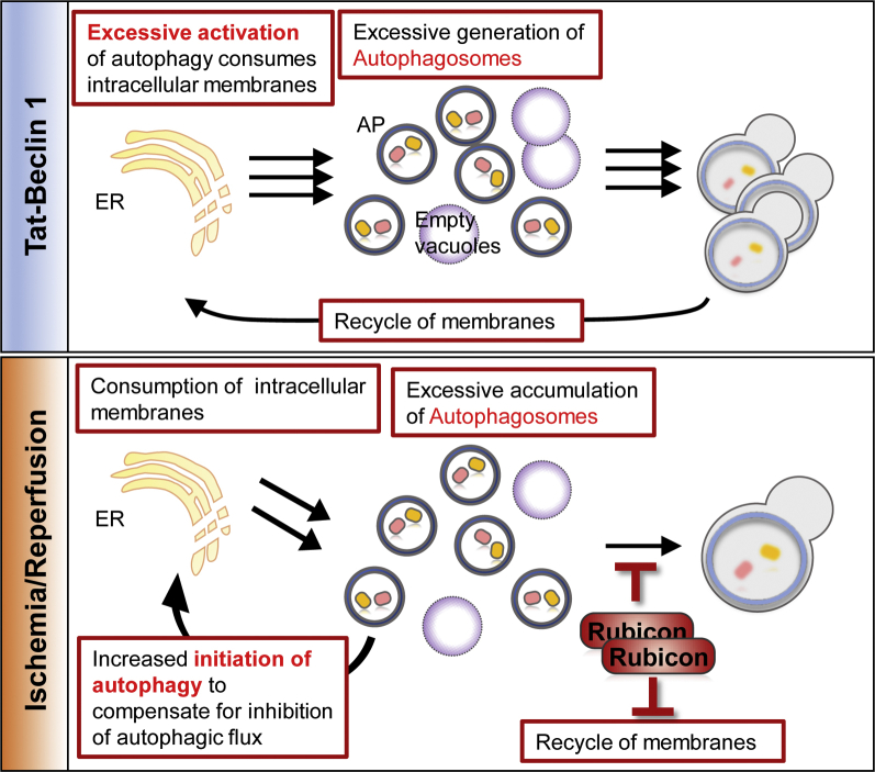
Excessive autophagy induces a defined form of cell death called autosis, which is characterized by unique morphological features, including ballooning of perinuclear space and biochemical features, including sensitivity to cardiac glycosides. Autosis is observed during the late phase of reperfusion after a period of ischemia and contributes to myocardial injury. This review discusses unique features of autosis, the involvement of autosis in myocardial injury, and the molecular mechanism of autosis. Because autosis promotes myocardial injury under some conditions, a better understanding of autosis may lead to development of novel interventions to protect the heart against myocardial stress.

Keywords: ATG, autophagy-related; ATPase, adenosine triphosphatase; ER, endoplasmic reticulum; HIV, human immunodeficiency virus; I/R, ischemia-reperfusion; LBR, lamin B receptor; Na+,K+–adenosine triphosphatase; PI3K, phosphatidylinositol 3 kinase; PNS, perinuclear space; Tat, transactivation of transcription; autophagic cell death; autophagic flux; autosis; beclin 1; rubicon.


Figures

None
Graphical abstract
Figure 1
Figure 1
Molecular Mechanism of Autophagy The process of autophagy comprises multiple steps, including initiation, nucleation, elongation and completion, and fusion and degradation. In the initiation step, the Unc-51-like kinase 1 (ULK1)/autophagy-related protein 1 (Atg1) complex is regulated by mammalian target of rapamycin (mTOR) inhibition in response to autophagy-inducing conditions, such as starvation. Thereafter, the ULK1/Atg1 complex activates the phosphatidylinositol 3 kinase complex to nucleate autophagosomal membranes. In the elongation step, 2 ubiquitin-like conjugation systems, the Atg12 and light chain 3 (LC3) conjugation systems, expand autophagosomes. After completion of elongation, autophagosomes fuse with lysosomes to degrade cargo materials. Rubicon associated with the phosphatidylinositol 3 kinase complex negatively regulates the fusion process. Atg13 = autophagy-related protein 13; Atg16L = autophagy-related protein 16 like; Bcl2 = B-cell lymphoma 2; FIP200 = FAK family kinase-interacting protein of 200 kD; mLST8 = mammalian lethal with SEC13 protein 8; p = phosphorylation; PE = phosphatidylethanolamine; UVRAG = ultraviolet radiation resistance–associated gene; Vps15 = vacuolar protein sorting 15; Vps34 = vacuolar protein sorting 34.
Figure 2
Figure 2
Morphological Features of Autosis During the early phase of autosis, the numbers of autophagosomes (APs), autolysosomes (ALs), and empty vacuoles (EVs) are drastically increased and separation of the inner and outer nuclear membranes is observed. The later phase is characterized by focal nuclear concavity, focal ballooning of the perinuclear space (PNS), and disappearance of subcellular organelles. ER = endoplasmic reticulum; Nu = nucleus.
Central Illustration
Biochemical Features of Autosis Autosis is activated by the autophagy-inducing peptide, tyrosine aminotransferase (Tat)–beclin 1, hypoxia-ischemia in neonatal cerebral brain and ischemia-reperfusion in the heart. Autosis is regulated by the physical interaction between beclin 1 and Na+,K+–adenosine triphosphatase (ATPase), which is inhibited by cardiac glycosides, by up-regulation of rubicon, which attenuates fusion of autophagosome and lysosome, and by the core autophagy machinery. Atg5 = autophagy-related protein 5.
Figure 3
Figure 3
Cause of Autotic Cell Death (Top) Excessive generation of autophagic vacuoles in response to tyrosine aminotransferase (Tat)–beclin 1 causes excessive consumption of intracellular membrane in cardiomyocytes in vitro. (Bottom) Accumulation of autophagic vacuoles induced by up-regulation of rubicon causes excessive consumption of intracellular membrane in cardiomyocytes in vivo. Abbreviations as in Figure 2.

References

    1. Pyo J.O., Nah J., Jung Y.K. Molecules and their functions in autophagy. Exp Mol Med. 2012;44:73–80. - PMC - PubMed
    1. Kroemer G., Levine B. Autophagic cell death: the story of a misnomer. Nat Rev Mol Cell Biol. 2008;9:1004–1010. - PMC - PubMed
    1. Matsui Y., Takagi H., Qu X. Distinct roles of autophagy in the heart during ischemia and reperfusion: roles of AMP-activated protein kinase and beclin 1 in mediating autophagy. Circ Res. 2007;100:914–922. - PubMed
    1. Zhu H., Tannous P., Johnstone J.L. Cardiac autophagy is a maladaptive response to hemodynamic stress. J Clin Invest. 2007;117:1782–1793. - PMC - PubMed
    1. Kobayashi S., Volden P., Timm D., Mao K., Xu X., Liang Q. Transcription factor GATA4 inhibits doxorubicin-induced autophagy and cardiomyocyte death. J Biol Chem. 2010;285:793–804. - PMC - PubMed

LinkOut - more resources