. 2020 Aug 12;5(33):21015-21023.
doi: 10.1021/acsomega.0c02580. eCollection 2020 Aug 25.

Thermosensitive Injectable Chitosan/Collagen/β-Glycerophosphate Composite Hydrogels for Enhancing Wound Healing by Encapsulating Mesenchymal Stem Cell Spheroids

Affiliations

Thermosensitive Injectable Chitosan/Collagen/β-Glycerophosphate Composite Hydrogels for Enhancing Wound Healing by Encapsulating Mesenchymal Stem Cell Spheroids

Ming Yang et al. ACS Omega. .

Abstract

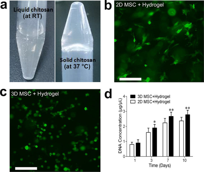
Chronic wounds caused by diabetic or venous diseases remain a social and healthcare burden. In this work, a new strategy is proposed in which injectable thermosensitive chitosan/collagen/β-glycerophosphate (β-GP) hydrogels were combined with three-dimensional mesenchymal stem cell (3D MSC) spheroids to accelerate chronic wound healing by enhanced vascularization and paracrine effects. Chitosan/collagen/β-GP solution mixed with 3D MSC spheroids was rapidly transformed to a gel at body temperature by physical cross-linking, then overlapped the wounds fully and fitted to any shape of the wound. The results showed that the combination therapy exhibited a markedly therapeutic effect than the hydrogel-loaded two-dimensional (2D) MSCs or 2D MSCs alone. The hydrogel could provide an environment conductive to the attachment and proliferation of encapsulated MSCs, especially accelerating the proliferation and paracrine factor secretion of 3D MSC spheroids. These results supplied a novel alternative approach to treat chronic wounds caused by diabetic or venous diseases.


Conflict of interest statement

The authors declare no competing financial interest.

Figures

Figure 1
Figure 1
Formation of 3D MSC spheroids and cell viability. (a) Hanging-drop method. Hanging drops, each containing approximately 5000 cells, are deposited and grown under a 10 cm dish lid. (b, c) Morphology of 3D MSC spheroids with size homogeneity. (d) Representative live/dead image showing cell viability of a 3D MSC spheroid. 3D MSC spheroids were stained with calcein-AM/ethidium homodimer-1 to visualize living (green) and dead (red) cells (d). (e) MSC spheroid diameters after culture for 36 and 60 h were measured with a particle analyzer using Image J software (measurement of 200 spheroids each). (f) Size distribution of MSC spheroids cultured for 60 h is shown. Scale bar in (b) was 200 μm, scale bars in (c, d) were 50 μm.
Figure 2
Figure 2
Thermosensitive property and cell proliferation in a 3D MSC spheroid-encapsulated chitosan/collagen/β-GP hydrogel. (a) Representative images of a chitosan/collagen/β-GP hydrogel showing thermosensitive property. The hydrogel changed from a liquid at room temperature (RT) to a gel at 37 °C. (b, c) Fluorescence microscopic images of calcein-AM staining of 2D MSC (b) and 3D MSC spheroids (c) encapsulated in a chitosan/collagen/β-GP hydrogel after 1 week of culture. Representative images are shown. Scale bar was 200 μm. (d) MSC proliferation in the hydrogel was determined by DNA content quantification at days 1, 3, 7, and 10. Experiments were performed in triplicate wells. Data are representative of three independent experiments (n = 3; *, P < 0.05; **, P < 0.01).
Figure 3
Figure 3
Wound-healing acceleration in 3D MSC spheroid-encapsulated chitosan/collagen/β-GP hydrogel-treated diabetic mice. (a) Representative images of the wound in db/db mice at week(s) 0, 1, 2, and 3. Murine excisional wound splintings (top panel) were created on the dorsal skin of db/db mice and the wound received implantation of 2D monolayer MSC without the hydrogel as a control, a 2D monolayer MSC-encapsulated chitosan/collagen/β-GP hydrogel, and a 3D MSC spheroid-encapsulated chitosan/collagen/β-GP hydrogel. The photographs of the first three panels were taken with transparent Tegaderm dressing, and those from the last panel were taken after removal of the dressing. (b) Wound closure areas were measured at week(s) 1, 2, and 3 in the control group (n = 12), the 2D MSC + hydrogel group (n = 10), and the 3D MSC + hydrogel group (n = 14). Analysis of variance (ANOVA): versus control, *, P < 0.05; **, P < 0.01; and versus 2D MSC + hydrogel group, ##, P < 0.01. Abbreviation: 2D, two-dimensional; 3D, three-dimensional; MSC, mesenchymal stem cells; wk, week(s).
Figure 4
Figure 4
Histological examination of the wound tissue in diabetic mice at week(s) 1 and 3 after implantation. (a) Representative 1 week-wound or 3 week-wound tissue samples stained with hematoxylin and eosin (H&E). Black arrows show arterioles containing erythrocytes. Scale bar was 500 μm. (b) Microphotographs of the wound bed from different transplantation groups at week 3 show the rate of re-epithelization and closure in the wound. Scale bar was 2 mm. Abbreviations: Ep, epidermis, wk, week.
Figure 5
Figure 5
Effects of the 3D MSC spheroid-encapsulated chitosan/collagen/β-GP hydrogel on the vascularization in the wound. (a) Representative confocal images of wound tissue sections immunostained with a vascular endothelial antibody CD31 2 weeks after transplantation. Nuclei (blue) were stained with 4,6-diamidino-2-phenylindole (DAPI). Scale bar = 50 μm. (b) Micrograph of microvessels in the 2-week-old wound area. The microvessel density within the wound area (×200) is higher in the 3D MSC spheroid-encapsulated chitosan/collagen/β-GP hydrogel-treated group compared with other two groups. (c) Densities of microvessels in wounds were assessed morphometrically after immunostaining for CD31 (n = 8). Analysis of variance (ANOVA); versus control, *, P < 0.05; **, P < 0.01; and versus 2D MSC + hydrogel group, #, P < 0.01.
Figure 6
Figure 6
Expression of paracrine cytokines in wound tissues. The differential expression of VEGFa, Ang1, CXCR4, and SDF1 protein in 2D monolayer MSC-treated (control), 2D monolayer MSC-encapsulated chitosan/collagen/β-GP hydrogel-treated, and 3D MSC spheroid-encapsulated chitosan/collagen/β-GP hydrogel-treated wounds were detected by western blot assay (a) and quantified (b) after the first week of treatment. The experiment was repeated three times, and one representative result is shown. Analysis of variance (ANOVA), versus control, *, P < 0.05; and versus 2D MSC + hydrogel group, #, P < 0.01. Abbreviations: Ang, angiopoietin; VEGF, vascular endothelial growth factor.

References

    1. Shaw T. J.; Martin P. Wound repair at a glance. J. Cell Sci. 2009, 122, 3209–3213. 10.1242/jcs.031187. - DOI - PMC - PubMed
    1. Singer A. J.; Clark R. A. Cutaneous wound healing. N. Engl. J. Med. 1999, 341, 738–746. 10.1056/NEJM199909023411006. - DOI - PubMed
    1. Falanga V. Wound healing and its impairment in the diabetic foot. Lancet 2005, 366, 1736–1743. 10.1016/S0140-6736(05)67700-8. - DOI - PubMed
    1. Martin P.; Nunan R. Cellular and molecular mechanisms of repair in acute and chronic wound healing. Br. J. Dermatol. 2015, 173, 370–378. 10.1111/bjd.13954. - DOI - PMC - PubMed
    1. Arno A. I.; Amini-Nik S.; Blit P. H.; Al-Shehab M.; Belo C.; Herer E.; Tien C. H.; Jeschke M. G. Human Wharton’s jelly mesenchymal stem cells promote skin wound healing through paracrine signaling. Stem Cell Res. Ther. 2014, 5, 28.10.1186/scrt417. - DOI - PMC - PubMed