. 2021 Jan;231(1):e13553.
doi: 10.1111/apha.13553. Epub 2020 Sep 17.

The body region specificity in murine models of muscle regeneration and atrophy

Affiliations

The body region specificity in murine models of muscle regeneration and atrophy

Kiyoshi Yoshioka et al. Acta Physiol (Oxf). 2021 Jan.

Abstract

Aim: Skeletal muscles are distributed throughout the body, presenting a variety of sizes, shapes and functions. Here, we examined whether muscle regeneration and atrophy occurred homogeneously throughout the body in mouse models.

Methods: Acute muscle regeneration was induced by a single intramuscular injection of cardiotoxin in adult mice. Chronic muscle regeneration was assessed in mdx mice. Muscle atrophy in different muscles was evaluated by cancer cachexia, ageing and castration mouse models.

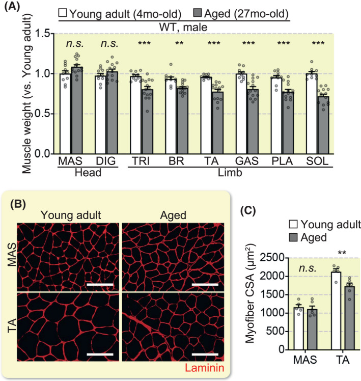
Results: We found that, in the cardiotoxin-injected acute muscle injury model, head muscles slowly regenerated, while limb muscles exhibited a rapid regeneration and even overgrowth. This overgrowth was also observed in limb muscles alone (but not in head muscles) in mdx mice as chronic injury models. We described the body region-specific decline in the muscle mass in muscle atrophy models: cancer cachexia-induced, aged and castrated mice. The positional identities, including gene expression profiles and hormone sensitivity, were robustly preserved in the ectopically engrafted satellite cell-derived muscles in the castrated model.

Conclusion: Our results indicate that positional identities in muscles should be considered for the development of efficient regenerative therapies for muscle weakness, such as muscular dystrophy and age-related sarcopenia.

Keywords: ageing; cancer cachexia; castration; heterogeneity; muscle atrophy; positional memory; skeletal muscle.


Conflict of interest statement

The authors declare that they have no competing interests.

Figures

FIGURE 1
FIGURE 1
Regional specificity in muscle regeneration. (A–E) Muscle regeneration was induced by cardiotoxin (CTX) injection into the masseter (MAS) and the tibialis anterior (TA) muscles of adult male mice. (A) Illustration of the experiment and representative images of muscle tissues 40 weeks after a CTX injection. Scale bars, 5 mm. (B) Time course of regenerating muscle weight change normalized to that of intact muscles (n = 4 mice, # P < .05, ## P < .01, ### P < .001, intact vs. CTX. ***P < .001, MAS vs. TA). (C) Number of myofibers per whole muscle tissue section 40 weeks post‐injury (n = 3 mice, **P < .01). (D) Mean cross‐sectional area (CSA) of intact (CTX non‐injected side) or centrally nucleated (CTX‐injected side) myofibers (n = 4 mice). (E) CSA distribution of MAS and TA at 40 weeks post‐injury (n = 4 mice). Scale bars, 100 µm. ‐ Data represent mean ± SEM. All error bars show the standard error of the mean (SEM)
FIGURE 2
FIGURE 2
Region‐specific muscle regeneration in mdx mice. (A) Illustration of muscles. SS, supraspinatus; TRI, triceps brachii; BR, brachioradialis; GAS, gastrocnemius; PLA, plantaris; SOL, soleus. (B) Representative images of MAS and TA tissues in wild‐type (WT) and mdx mice. Scale bars, 5 mm. (C) Muscle weights in mdx mice normalized to those in WT mice (WT; n = 8 mice, mdx; n = 8 mice). (D) Mean CSA of regenerative myofibers (WT; n = 5 mice, mdx; n = 6 mice). (E) CSA distribution of MAS and TA (WT; n = 5, mdx; n = 6). Scale bars, 100 µm. Data represent mean ± SEM. All error bars show the standard error of the mean (SEM). **P < .01, ***P < .001. MAS, masseter; DIG, digastric; SS, supraspinatus; TRI, triceps brachii; BR, brachioradialis; TA, tibialis anterior; EDL, extensor digitorum longus; GAS, gastrocnemius; PLA, plantaris; SOL, soleus
FIGURE 3
FIGURE 3
Region‐specific phenotypes in cancer cachexia. (A) Cancer cachexia was induced by an LLC injection to induce systemic muscle atrophy. (B) Body weight and epididymal fat were weighed (CON; n = 5 mice, LLC; n = 5 mice). (C) Muscle weight (LLC/ CON) at 4 weeks following an LLC injection (CON; n = 8 mice, LLC; n = 7 mice). FDP: flexor digitorum profundus. (D) Representative images of HE‐stained muscle tissue sections. Mean myofiber CSA (MAS, FDP, PLA and SOL; n = 5 mice, TA; n = 4 mice). Scale bars, 100 µm. (E) Expression of the atrophy‐related genes in MAS and TA (n = 5 mice each). All error bars show the SEM. *P < .05, **P < .01, ***P < .001. MAS, masseter; DIG; TRI, triceps brachii; BR, brachioradialis; FDP, flexor digitorum profundus; TA, tibialis anterior; GAS, gastrocnemius; PLA, plantaris; SOL, soleus
FIGURE 4
FIGURE 4
Impact of ageing on head and limb muscles. (A) Muscle weight ratio (young adult; n = 10 mice, aged; n = 13 mice). (B) Representative immunohistochemical images of MAS and TA tissue sections stained for laminin. (C) Mean myofiber CSA (young adult; n = 5 mice, aged; n = 6 mice). Scale bars, 100 µm. All error bars show the SEM. **P < .01, ***P < .001. MAS, masseter; DIG; TRI, triceps brachii; BR, brachioradialis; TA, tibialis anterior; GAS, gastrocnemius; PLA, plantaris; SOL, soleus
FIGURE 5
FIGURE 5
Region‐specific effects of castration on muscles. (A) Castration was conducted using an androgen insufficient model. (B) Representative muscle tissue images of the LA and BUL. (C) Muscle weight ratio (sham/castration) 2 weeks after the operation (n = 5 mice each). (D) Representative phase‐contrast images of individual myofibers isolated from EDL and US. (E) The diameter of individual myofibers (n = 4 mice each, 30 myofibers were counted per mouse). Scale bars, 50 µm. All error bars show the SEM. *P < .05, **P < .01, ***P < .001. MAS, masseter; DIG, digastric; SS, supraspinatus; TRI, triceps brachii; BR, brachioradialis; TA, tibialis anterior; EDL, extensor digitorum longus; GAS, gastrocnemius; PLA, plantaris; SOL, soleus; LA, levator ani; BUL, bulbospongiosus; US, urethral sphincter
FIGURE 6
FIGURE 6
Positional identities in ectopically engrafted satellite cell‐derived myofibers. (A) Schematic illustration of engraftment. TA (Host: mdx mice) was irradiated and pre‐injured with a CTX injection. Satellite cells from the limb muscle or perineal muscle (Donor: Pax7‐YFP mice) were engrafted into the TA. (B) qPCR analysis of gene expression in TA and BUL (n = 6 mice each). (C) qPCR analysis of gene expression in the engrafted TA (n = 4 mice each). (D) Representative immunohistochemical image of transverse sections of TA for dystrophin. Scale bars, 100 µm. (E) Mean CSA of the 50 largest dystrophin‐positive myofibers in a section (n = 4 mice each). Two‐way ANOVA results were as follow. Donor SCs, **. Host mice, ***. Interaction, **. All error bars show the SEM. *P < .05, **P < .01, ***P < .001. BUL, bulbospongiosus; TA, tibialis anterior

References

    1. Schiaffino S, Reggiani C. Fiber types in mammalian skeletal muscles. Physiol Rev. 2011;91(4):1447‐1531. - PubMed
    1. Uchitomi R, Hatazawa Y, Senoo N, et al. Metabolomic Analysis of Skeletal Muscle in Aged Mice. Sci Rep. 2019;9(1):10425. - PMC - PubMed
    1. Mauro A. Satellite cell of skeletal muscle fibers. J Biophys Biochem Cytol. 1961;9:493‐495. - PMC - PubMed
    1. Morgan JE, Zammit PS. Direct effects of the pathogenic mutation on satellite cell function in muscular dystrophy. Exp Cell Res. 2010;316(18):3100‐3108. - PubMed
    1. Randolph ME, Pavlath GK. A muscle stem cell for every muscle: variability of satellite cell biology among different muscle groups. Front Aging Neurosci. 2015;7:190. - PMC - PubMed

Publication types