. 2020 Sep 1;76(Pt 9):889-898.
doi: 10.1107/S2059798320010505. Epub 2020 Aug 17.

Crystal structures of human ENPP1 in apo and bound forms

Affiliations

Crystal structures of human ENPP1 in apo and bound forms

Matthew L Dennis et al. Acta Crystallogr D Struct Biol. .

Abstract

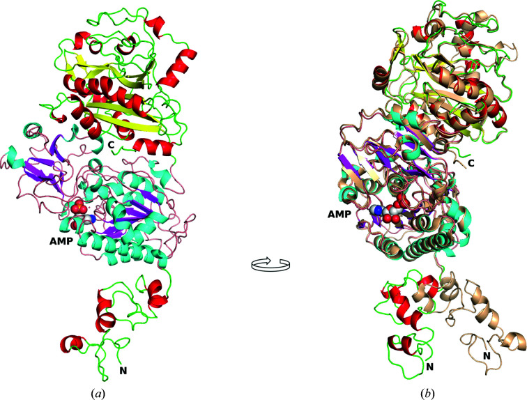
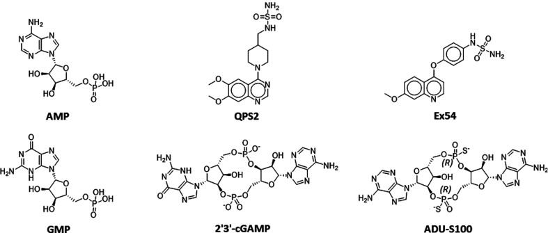
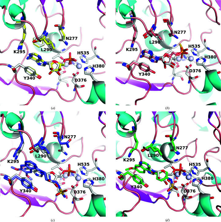
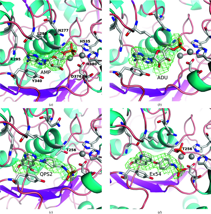
Cancer is one of the leading causes of mortality in humans, and recent work has focused on the area of immuno-oncology, in which the immune system is used to specifically target cancerous cells. Ectonucleotide pyrophosphatase/phosphodiesterase 1 (ENPP1) is an emerging therapeutic target in human cancers owing to its role in degrading cyclic GMP-AMP (cGAMP), an agonist of the stimulator of interferon genes (STING). The available structures of ENPP1 are of the mouse enzyme, and no structures are available with anything other than native nucleotides. Here, the first X-ray crystal structures of the human ENPP1 enzyme in an apo form, with bound nucleotides and with two known inhibitors are presented. The availability of these structures and a robust crystallization system will allow the development of structure-based drug-design campaigns against this attractive cancer therapeutic target.

Keywords: ENPP1; cancer; inhibitor structures; pyrophosphatase/phosphodiesterases.


Figures

Figure 1
Figure 1
Structure of hENPP1 with AMP and comparison with mouse ENPP1 (PDB entry 4b56). (a) The SMB domain is at the N-terminus and is shown with red helices, the catalytic domain is shown with cyan helices and magenta β-strands with AMP represented as spheres, and the nuclease-like domain is at the top of the figure and is shown with red helices and yellow β-strands. (b) The image is rotated approximately 90° clockwise and mouse ENPP1 (PDB entry 4b56) is superposed on the human structure. The mouse structure is coloured monochrome wheat with the N- and C-termini labelled. This figure was generated using PyMOL and GIMP.
Figure 2
Figure 2
Chemical structures of the compounds. The structures of AMP, GMP and cGAMP and the synthesized compounds ADU-S100, QPS2 and Ex54 are shown.
Figure 3
Figure 3
Relationship of structures with bound AMP. (a) The human and mouse (PDB entry 4gtw) structures of ENPP1 bound to AMP. hENPP1 has grey C atoms and mENPP1 has yellow C atoms for both the AMP molecules and the surrounding residues highlighted in stick representation (Asn277, Leu290, Lys295 and Trp340). The two Zn atoms are shown as grey spheres and the active-site residues are also highlighted in stick representation (three of these, Asp376, His380 and His535, are labelled). (b) hENPP1 with AMP superposed with the hydrolysed product of ADU-S100, with hENPP1 with AMP using the same colour scheme as in (a) and ADU-S100 coloured burnt orange. (c) hENPP1 with AMP superposed with hENPP1 with QPS2 (purple C atoms). (d) hENPP1 with AMP superposed hENPP1 with Ex54 (green C atoms). This figure was generated using PyMOL and GIMP.
Figure 4
Figure 4
OMIT maps of compounds bound to hENPP1. 3σ difference OMIT maps of AMP (a), ADU-S100 (b), QPS2 (c) and Ex54 (d). The orientation is approximately the same as that in Fig. 3 ▸, with the Zn atoms shown as grey spheres and various residues in the binding site highlighted in stick representation. Note that Ex54 is bound such that Thr256 is still phosphorylated. This figure was generated with PyMOL and GIMP.
Figure 5
Figure 5
Superposition of Ex54 and QPS2 on AMP in hENPP1. AMP has green C atoms, Ex54 has cyan C atons and QPS2 has magenta C atoms. The Zn atoms of the active site are shown as grey spheres and the two water molecules found in the AMP-bound structure but not in the inhibitor-bound structures are shown as red spheres. The methoxy moieties of the inhibitors occupy approximately the same space as that occupied by the two waters in the AMP-bound structure. (a) and (b) show two different orientations to highlight that the fused rings are all in the same plane and that the phosphate moiety of AMP is found in a different orientation to that of the sulfamides of the two inhibitors.

References

    1. Ablasser, A., Schmid-Burgk, J. L., Hemmerling, I., Horvath, G. L., Schmidt, T., Latz, E. & Hornung, V. (2013). Nature, 503, 530–534. - PMC - PubMed
    1. Aerts, I., Martin, J.-J., De Deyn, P. P., Van Ginniken, C., Van Ostade, X., Kockx, M., Dua, G. & Slegers, H. (2011). Clin. Neurol. Neurosurg. 113, 224–229. - PubMed
    1. Carozza, J. A., Böhnert, V., Nguyen, K. C., Skariah, G., Shaw, K. E., Brown, J. A., Rafat, M., von Eyben, R., Graves, E. E., Glenn, J. S., Smith, M. & Li, L. (2020). Nat. Cancer, 1, 184–196. - PMC - PubMed
    1. Corrales, L., Glickman, L. H., McWhirter, S. M., Kanne, D. B., Sivick, K. E., Katibah, G. E., Woo, S.-R., Lemmens, E., Banda, T., Leong, J. J., Metchette, K., Dubensky, T. W. & Gajewski, T. F. (2015). Cell. Rep. 11, 1018–1030. - PMC - PubMed
    1. Dimatteo, C., Marucci, A., Palazzo, A., Cisternino, C., Marsano, R. M., Trischitta, V. & Di Paola, R. (2013). Biochim. Biophys. Acta, 1833, 552–558. - PubMed