. 2020 Sep 2:9:e57132.
doi: 10.7554/eLife.57132.

CD163 and pAPN double-knockout pigs are resistant to PRRSV and TGEV and exhibit decreased susceptibility to PDCoV while maintaining normal production performance

Affiliations

CD163 and pAPN double-knockout pigs are resistant to PRRSV and TGEV and exhibit decreased susceptibility to PDCoV while maintaining normal production performance

Kui Xu et al. Elife. .

Abstract

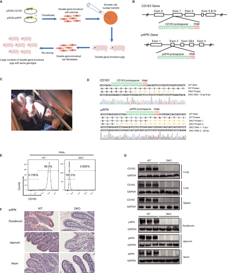
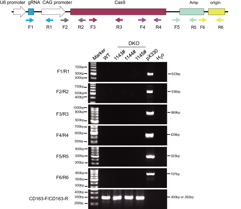
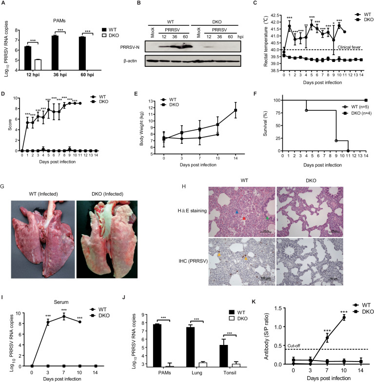
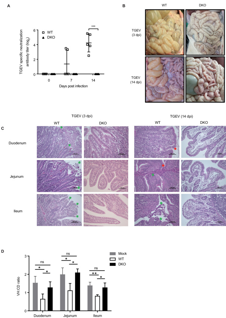
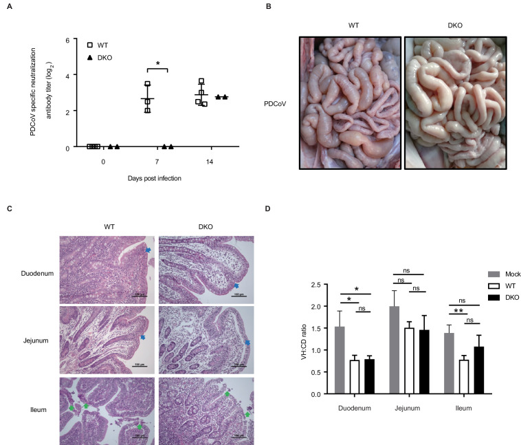
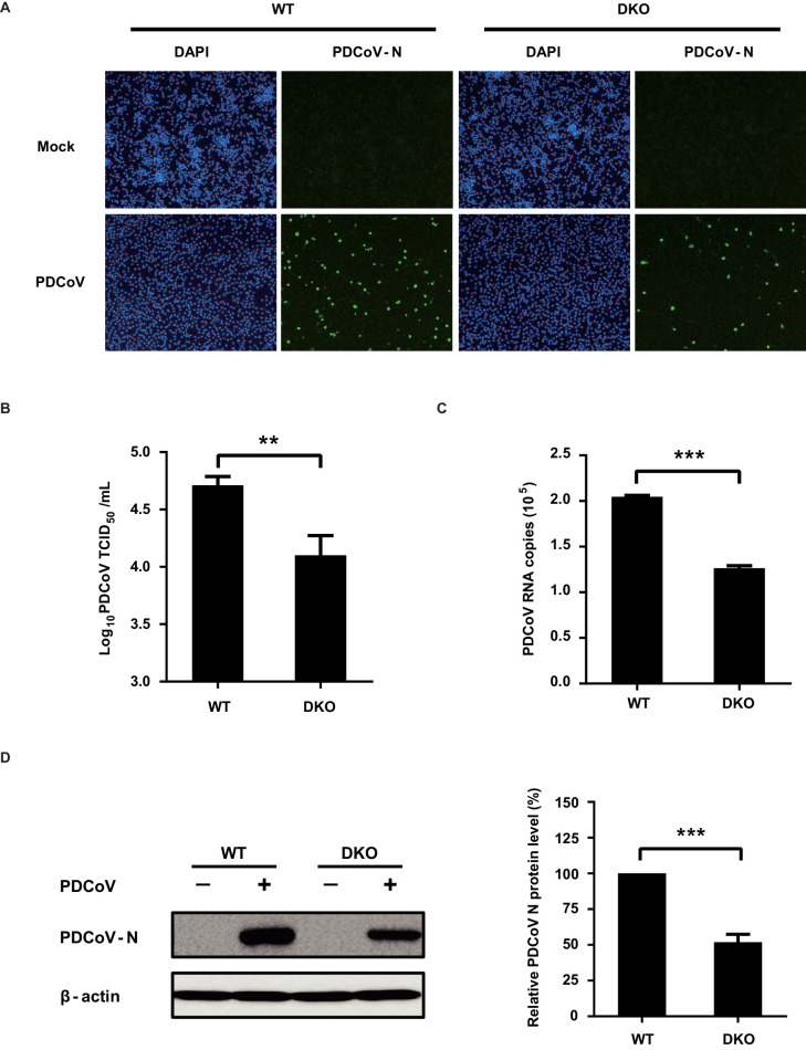
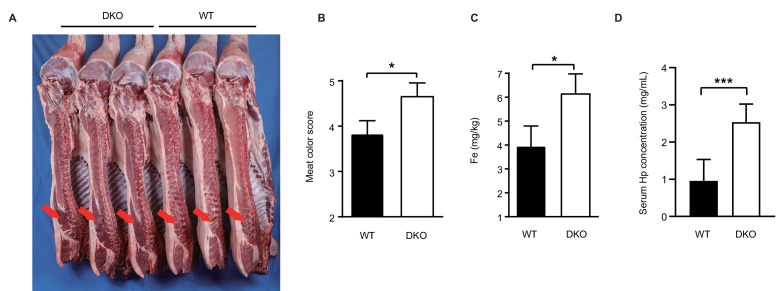
Porcine reproductive and respiratory syndrome virus (PRRSV) and transmissible gastroenteritis virus (TGEV) are two highly infectious and lethal viruses causing major economic losses to pig production. Here, we report generation of double-gene-knockout (DKO) pigs harboring edited knockout alleles for known receptor proteins CD163 and pAPN and show that DKO pigs are completely resistant to genotype 2 PRRSV and TGEV. We found no differences in meat-production or reproductive-performance traits between wild-type and DKO pigs, but detected increased iron in DKO muscle. Additional infection challenge experiments showed that DKO pigs exhibited decreased susceptibility to porcine deltacoronavirus (PDCoV), thus offering unprecedented in vivo evidence of pAPN as one of PDCoV receptors. Beyond showing that multiple gene edits can be combined in a livestock animal to achieve simultaneous resistance to two major viruses, our study introduces a valuable model for investigating infection mechanisms of porcine pathogenic viruses that exploit pAPN or CD163 for entry.

Keywords: CRISPR/Cas9; PDCoV; PRRSV; TGEV; infectious disease; microbiology; pig; virus.

Plain language summary

Pig epidemics are the biggest threat to the pork industry. In 2019 alone, hundreds of billions of dollars worldwide were lost due to various pig diseases, many of them caused by viruses. The porcine reproductive and respiratory virus (PRRS virus for short), for instance, leads to reproductive disorders such as stillbirths and premature labor. Two coronaviruses – the transmissible gastroenteritis virus (or TGEV) and the porcine delta coronavirus – cause deadly diarrhea and could potentially cross over into humans. Unfortunately, there are still no safe and effective methods to prevent or control these pig illnesses, but growing disease-resistant pigs could reduce both financial and animal losses. Traditionally, breeding pigs to have a particular trait is a slow process that can take many years. But with gene editing technology, it is possible to change or remove specific genes in a single generation of animals. When viruses infect a host, they use certain proteins on the surface of the host’s cells to find their inside: the PRRS virus relies a protein called CD163, and TGEV uses pAPN. Xu, Zhou, Mu et al. used gene editing technology to delete the genes that encode the CD163 and pAPN proteins in pigs. When the animals were infected with PRRS virus or TGEV, the non-edited pigs got sick but the gene-edited animals remained healthy. Unexpectedly, pigs without CD163 and pAPN also coped better with porcine delta coronavirus infections, suggesting that CD163 and pAPN may also help this coronavirus infect cells. Finally, the gene-edited pigs reproduced and produced meat as well as the control pigs. These experiments show that gene editing can be a powerful technology for producing animals with desirable traits. The gene-edited pigs also provide new knowledge about how porcine viruses infect pigs, and may offer a starting point to breed disease-resistant animals on a larger scale.


Conflict of interest statement

KX, YZ, YM, ZL, SH, YX, LF, YW, XZ, CX, JC, ZF, GX, JG, HL, SX, JL, KL No competing interests declared, CG is affiliated with Shandong Landsee Genetics Co., Ltd. The author has no financial interests to declare. HS is affiliated with Shenzhen Kingsino Technology Co., Ltd. The author has no financial interests to declare.

Figures

Figure 1.
Figure 1.. Generation of CD163 and pAPN DKO pigs by CRISPR/Cas9.
(A) Schematic overview of the generation process for DKO cloned pigs. (B) Genetic maps of the CD163 (top) and pAPN (bottom) genes with the location and sequences of the sgRNAs. Exons, white boxes; sgRNA protospacer sequences, green; and PAM sequences, red. (C) Four F0 generation cloned pigs (1085#, 1143#, 1144#, 1145#) aged 1-month-old and a surrogate. (D) Sanger sequencing confirmation of DKO genotypes for three F0 cloned piglets (1143#, 1144#, 1145#). sgRNA protospacer sequences, green; PAM sequences, red; predicted amino acid sequences resulting from frameshift mutations, yellow. (E) Detection of CD163 expression on the surface of PAMs by flow cytometry. (F) Detection of pAPN expression in different small intestine segments by IHC. PAMs and tissues were derived from DKO and WT pigs. (G) Western blot analysis confirmed CD163 and pAPN expression are undetectable in different tissues of DKO pigs (N = 3).
Figure 1—figure supplement 1.
Figure 1—figure supplement 1.. PCR detection of random integration of the pX330 plasmid backbone.
Different colored and shaped boxes represent elements on the pX330 plasmid, arrows behind the boxes show the binding sites of primers in pX330 plasmid. Primer sequences are listed in Supplementary file 6.
Figure 2.
Figure 2.. DKO pigs are resistant to genotype 2 PRRSV infection.
(A) qRT-PCR detection of PRRSV load and (B) western blot confirmation of PRRSV-N protein expression in PAMs from WT and DKO pigs at 12, 36, and 60 hpi (MOI = 0.1). (C) Rectal temperatures and (D) clinical symptoms scores were recorded daily beginning at 0 dpi to 14 dpi. Temperatures > 40°C were considered fever. WT: 0 to 4 dpi, N = 6; 5 to 8 dpi, N = 5; 9 dpi, N = 4; 10 dpi, N = 2; 11 dpi, N = 1. DKO: 0 to 14 dpi, N = 4. (E) Body weights measured at 0, 3, 7, 10, and 14 dpi. WT: 0 to 3 dpi, N = 6; 7 dpi, N = 5; 10 dpi, N = 2. DKO: 0 to 14 dpi, N = 4. (F) Survival curves of WT and DKO pigs with PRRSV. WT, N = 5; DKO, N = 4. (G) Representative lesions of infected lungs. (H) H and E staining (top) of lesions in lung tissue; IHC (bottom) detection of viral antigens in PRRSV-infected lungs. Lymphocytic infiltration (blue triangle); necrotic cells in the alveolar wall (red triangle); bronchial wall dilated and filled with serous fluid (green triangle); PRRSV-N protein (yellow triangle) (I) PRRSV loads in serum at 0, 3, 7, 10, and 14 dpi. WT: 0 to 3 dpi, N = 6; 7 dpi, N = 5; 10 dpi, N = 2. DKO: 0 to 14 dpi, N = 4. (J) PRRSV loads in PAMs (WT group: N = 3; DKO group: N = 3), lung tissues (WT: N = 5; DKO: N = 3), and tonsil tissues (WT: N = 5; DKO: N = 4). (K) PRRSV-specific antibodies in serum. WT: 0 to 3 dpi, N = 6; 7 dpi, N = 5; 10 dpi, N = 2. DKO: 0 to 14 dpi, N = 4. Data are expressed as means ± SD. Statistical significance was determined by Student’s t test; ns, p>0.05; *p<0.05; **p<0.01; ***p<0.001.
Figure 3.
Figure 3.. DKO pigs are resistant to TGEV infection.
(A) TGEV-specific antibody detection in serum of WT and DKO pigs at 0, 7, and 14 dpi. WT group: N = 5; DKO group: N = 4. (B) Representative macroscopic lesion of small intestines at 3 dpi (top) and 14 dpi (bottom) in WT and DKO pigs. (C) H and E staining of lesions small intestine sections derived from both WT and DKO pigs at 3 dpi (left) and 14 dpi (right). H and E staining shows intestinal villi fusion, plasma cells accumulating in the lamina propria, and infiltration of eosinophils (red arrow), and necrosis and shedding of intestinal mucosal epithelial cells (green arrow). (D) The ratio of intestinal villus height to the crypt depth derived from both WT and DKO pigs at 3 dpi. Mock: duodenum to ileum, N = 3. WT: duodenum to ileum, N = 3. DKO: duodenum, N = 4; jejunum, N = 3; ileum, N = 4. Data are expressed as the mean ± SD. Statistical significance was determined by Student’s t-test; ns, p>0.05; *p<0.05; **p<0.01; ***p<0.001.
Figure 4.
Figure 4.. DKO pigs exhibit reduced susceptibility to PDCoV.
(A) PDCoV-specific antibody detection in serum from both WT and DKO pigs at 0, 7, and 14 dpi. WT group: 0 dpi, N = 4; 7 dpi, N = 3; 14 dpi, N = 4. DKO group, N = 2. (B) Representative macroscopic lesions of the small intestines at 14 dpi from WT and DKO groups of pigs challenged with PDCoV. (C) H and E staining of small intestine segments sections to detect lesions; small intestine tissues were derived from PDCoV-infected WT and DKO pigs. Intestinal villi fusion, infiltration of lymphocytes in the intestinal mucosa, and a large number in the intrinsic membrane (blue arrow), and necrosis and shedding of intestinal mucosal intraepithelial cells and naked lamina propria (green arrow). (D) The ratio of intestinal villus height to the crypt depth derived from both WT and DKO pigs at 14 dpi. Mock: duodenum to ileum, N = 3. WT: duodenum to ileum, N = 3. DKO: duodenum to ileum, N = 3. Data are expressed as the mean ± SD. Statistical significance was determined by Student’s t-test; ns, p>0.05; *p<0.05; **p<0.01; ***p<0.001.
Figure 4—figure supplement 1.
Figure 4—figure supplement 1.. PAMs of DKO pigs exhibit reduced susceptibility to PDCoV.
(A) WT PAMs and DKO PAMs were infected or mock-infected with PDCoV (MOI = 10); at 24 hpi, PDCoV-N-specific fluorescence signals were detected by IFA. (B) WT PAMs and DKO PAMs were infected with PDCoV (MOI = 10); at 24 hpi, cells were collected and the viral titer was determined by TCID50 assays (in LLC-PK1 cells). (C, D) WT PAMs and DKO PAMs were infected with PDCoV (MOI = 10); at 24 hpi, cells were harvested and analyzed using qRT-PCR (C) and western blot assays (D). The PDCoV-N protein level was quantified by ImageJ. Data are expressed as the mean ± SD. Statistical significance was determined by Student’s t test; ns, p>0.05; *p<0.05; **p<0.01; ***p<0.001.
Figure 5.
Figure 5.. DKO pigs maintain normal production performance.
(A) Carcass photo of DKO and WT pigs. The meat of DKO pigs becomes darker red (red arrow). (B) Meat color scores for WT and DKO pigs. WT group: N = 3; DKO group: N = 3. (C) Detection of Fe content in longissimus dorsi muscle from both experimental groups. WT group: N = 3; DKO group: N = 3. (D) Serum Hp levels of WT and DKO pigs. WT group: N = 22; DKO group: N = 6. Data are expressed as the mean ± SD. Statistical significance was determined by Student’s t-test; ns, p>0.05; *p<0.05; **p<0.01; ***p<0.001.

References

    1. An K, Fang L, Luo R, Wang D, Xie L, Yang J, Chen H, Xiao S. Quantitative proteomic analysis reveals that transmissible gastroenteritis virus activates the JAK-STAT1 signaling pathway. Journal of Proteome Research. 2014;13:5376–5390. doi: 10.1021/pr500173p. - DOI - PubMed
    1. Brierley I, Digard P, Inglis SC. Characterization of an efficient coronavirus ribosomal frameshifting signal: requirement for an RNA pseudoknot. Cell. 1989;57:537–547. doi: 10.1016/0092-8674(89)90124-4. - DOI - PMC - PubMed
    1. Burkard C, Lillico SG, Reid E, Jackson B, Mileham AJ, Ait-Ali T, Whitelaw CB, Archibald AL. Precision engineering for PRRSV resistance in pigs: macrophages from genome edited pigs lacking CD163 SRCR5 domain are fully resistant to both PRRSV genotypes while maintaining biological function. PLOS Pathogens. 2017;13:e1006206. doi: 10.1371/journal.ppat.1006206. - DOI - PMC - PubMed
    1. Calvert JG, Slade DE, Shields SL, Jolie R, Mannan RM, Ankenbauer RG, Welch SK. CD163 expression confers susceptibility to porcine reproductive and respiratory syndrome viruses. Journal of Virology. 2007;81:7371–7379. doi: 10.1128/JVI.00513-07. - DOI - PMC - PubMed
    1. Chen J, Wang H, Bai J, Liu W, Liu X, Yu D, Feng T, Sun Z, Zhang L, Ma L, Hu Y, Zou Y, Tan T, Zhong J, Hu M, Bai X, Pan D, Xing Y, Zhao Y, Tian K, Hu X, Li N. Generation of pigs resistant to highly Pathogenic-Porcine reproductive and respiratory syndrome virus through gene editing of CD163. International Journal of Biological Sciences. 2019;15:481–492. doi: 10.7150/ijbs.25862. - DOI - PMC - PubMed

Publication types

MeSH terms