

A comparison of clinical, laboratory and chest CT findings of laboratory-confirmed and clinically diagnosed COVID-19 patients at first admission
- PMID: 32876570
- PMCID: PMC8136532
- DOI: 10.5152/dir.2020.20270
A comparison of clinical, laboratory and chest CT findings of laboratory-confirmed and clinically diagnosed COVID-19 patients at first admission
Abstract
Purpose: This study aims to identify chest computed tomography (CT) characteristics of coronavirus disease 2019 (COVID-19), investigate the association between CT findings and laboratory or demographic findings, and compare the accuracy of chest CT with reverse transcription-polymerase chain reaction (RT-PCR).
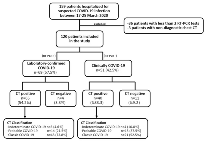
Methods: Overall, 120 of 159 consecutive cases isolated due to suspected COVID-19 at our hospital between 17 and 25 March 2020 were included in this retrospective study. All patients underwent both chest CT and RT-PCR at first admission. The patients were divided into two groups: laboratory-confirmed COVID-19 and clinically diagnosed COVID-19. Clinical findings, laboratory findings, radiologic features and CT severity index (CT-SI) of the patients were noted. The sensitivity, specificity, positive predictive value (PPV), negative predictive value (NPV), and accuracy of chest CT were calculated for the diagnosis of COVID-19, using RT-PCR as reference.
Results: The laboratory-confirmed and clinically diagnosed COVID-19 groups consisted of 69 (M/F 43/26, mean age 50.9±14.0 years) and 51 patients (M/F 24/27, mean age 50.9±18.8 years), respectively. Dry cough (62.3% vs. 52.9%), fever (30.4% vs. 25.5%) and dyspnea (23.2% vs. 27.5%) were the most common admission symptoms in the laboratory-confirmed and clinically diagnosed COVID-19 groups, respectively. Bilateral multilobe involvement (83.1% vs. 57.5%), peripheral distribution (96.9% vs. 97.5%), patchy shape (75.4% vs. 70.0%), ground-glass opacities (GGO) (96.9% vs. 100.0%), vascular enlargement (56.9% vs. 50.0%), intralobular reticular density (40.0% vs. 40.0%) and bronchial wall thickening (27.7% vs. 45.0%) were the most common CT findings in the laboratory-confirmed and clinically diagnosed COVID-19 subgroups, respectively. Except for the bilateral involvement and white blood cell (WBC) count, no difference was found between the clinical, laboratory, and parenchymal findings of the two groups. Positive correlation was found between CT-SI and, lactate dehydrogenase (LDH) and C-reactive protein (CRP) values in the laboratory-confirmed COVID-19 subgroup. Chest CT and RT-PCR positivity rates among patients with suspected COVID-19 were 87.5% (105/120) and 57.5% (69/120), respectively. The sensitivity, specificity, PPV, NPV and accuracy rates of chest CT were determined as 94.2% (95% confidence interval [CI], 85.8-98.4), 21.57% (95% CI, 11.3-35.3), 61.90% (95% CI, 58.2-65.5), 73.3% (95% CI, 48.2-89.1) and 63.3% (95% CI, 54.1-71.9), respectively.
Conclusion: Chest CT has high sensitivity and low specificity in the diagnosis of COVID-19. The clinical, laboratory, and CT findings of laboratory-confirmed and clinically diagnosed COVID-19 patients are similar.
Conflict of interest statement
The authors declared no conflicts of interest.
Figures
References
-
- Turkish Ministry of Health. COVID-19 (SARS-CoV-2 INFECTION) GUIDE-Study of Scientific Board. Turkish Ministry of Health; Ankara, Turkey: https://covid19.saglik.gov.tr/
-
- World Health Organization. Coronavirus disease 2019 (COVID-19) situation report–51. World Health Organization; Geneva: 2020. https://www.who.int/docs/default-source/coronaviruse/situation-reports/2....
Publication types
MeSH terms
LinkOut - more resources
Full Text Sources
Other Literature Sources
Medical
Research Materials
Miscellaneous
