Diagnostic performance of low-dose chest CT to detect COVID-19: A Turkish population study
- PMID: 32876571
- PMCID: PMC7963387
- DOI: 10.5152/dir.2020.20350
Diagnostic performance of low-dose chest CT to detect COVID-19: A Turkish population study
Abstract
Purpose: We aimed to evaluate the diagnostic performance of low-dose chest computed tomography (CT) in patients under investigation for coronavirus disease 2019 (COVID-19).
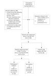
Methods: This retrospective study included 330 patients suspected of having COVID-19 from March 15 to April 16, 2020. We examined 306 patients upon initial presentation using both CT and real-time reverse-transcriptase polymerase-chain-reaction (rRT-PCR). The diagnostic performance of CT was calculated using rRT-PCR as a reference. Clinical and laboratory data, CT characteristics, and lesion distribution were assessed for patients with a confirmed diagnosis via rRT-PCR.
Results: A total of 250 patients were finally diagnosed with COVID-19. Clinical and laboratory findings included myalgia or fatigue (76%), fever (64.8%), dry cough (60.8%), elevated levels of C-reactive protein (86.4%), procalcitonin (62%), and D-dimer (58.2%), increased neutrophil-lymphocyte ratio (NLR) (54.8%), and lymphopenia (34%). Sensitivity, specificity, positive predictive value (PPV) and negative predictive value (NPV) of the initial CT scan were 90.4% (95% IC, 86%-93%), 64.2% (95% IC, 50%-76%), 91.8% (95% IC, 88%-94%), and 60% (95% IC, 49%-69%), respectively. The percentage of patients diagnosed on the initial rRT-PCR test was 51.6% (n=129). Most frequent CT characteristics of COVID-19 in the subgroup of rRT-PCR-positive patients were multiple lesion (97.4%, n=220), followed by bilateral involvement (88.5%, n=200), peripheral distribution (74.3%, n=168), ground-glass opacity (GGO) (69.2%, n=157), subpleural curvilinear opacity (41.6%, n=104), and mixed GGOs (27.6%, n=67).
Conclusion: rRT-PCR may produce initial false negative results. For this reason, typical CT findings for COVID-19 should be known especially by radiologists. We suggest that patients with typical CT findings but negative rRT-PCR results should be isolated, and rRT-PCR should be repeated to avoid misdiagnosis.
Conflict of interest statement
The authors declared no conflicts of interest.
Figures


References
-
- World Health Organization. Pneumonia of unknown cause: China. World Health Organization; Geneva: [Accessed February 13th, 2020]. Available via www.who.int/csr/don/05-january-2020-pneumonia-of-unkown-cause-china/en/
-
- World Health Organization main website. [Accessed March 12th, 2020]. https://www.who.int.
MeSH terms
LinkOut - more resources
Full Text Sources
Other Literature Sources
Medical
Research Materials
Miscellaneous

