. 2020 Dec;57(12):5299-5306.
doi: 10.1007/s12035-020-02105-y. Epub 2020 Sep 2.

An Experimental Model of Neurodegenerative Disease Based on Porcine Hemagglutinating Encephalomyelitis Virus-Related Lysosomal Abnormalities

Affiliations

An Experimental Model of Neurodegenerative Disease Based on Porcine Hemagglutinating Encephalomyelitis Virus-Related Lysosomal Abnormalities

Yungang Lan et al. Mol Neurobiol. 2020 Dec.

Abstract

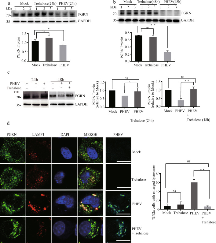
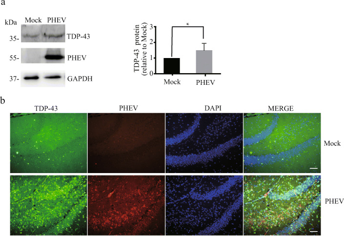
Lysosomes are involved in pathogenesis of a variety of neurodegenerative diseases and play a large role in neurodegenerative disorders caused by virus infection. However, whether virus-infected cells or animals can be used as experimental models of neurodegeneration in humans based on virus-related lysosomal dysfunction remain unclear. Porcine hemagglutinating encephalomyelitis virus displays neurotropism in mice, and neural cells are its targets for viral progression. PHEV infection was confirmed to be a risk factor for neurodegenerative diseases in the present. The findings demonstrated for the first time that PHEV infection can lead to lysosome disorders and showed that the specific mechanism of lysosome dysfunction is related to PGRN expression deficiency and indicated similar pathogenesis compared with human neurodegenerative diseases upon PHEV infection. Trehalose can also increase progranulin expression and rescue abnormalities in lysosomal structure in PHEV-infected cells. In conclusion, these results suggest that PHEV probably serve as a disease model for studying the pathogenic mechanisms and prevention of other degenerative diseases.

Keywords: Lysosomal abnormalities; Neurodegenerative diseases; Porcine hemagglutinating encephalomyelitis virus; Progranulin; Trehalose.


Conflict of interest statement

The authors declare that they have no conflict of interest.

Figures

Fig. 1
Fig. 1
PHEV infection leads to lysosomal abnormalities. a PHEV localizes to lysosomes, and PHEV infection results in greater lysosome enlargement compared with mock infection. The percentage of N2a cells containing enlarged lysosomes (> 1 μm) was quantified in the experiment. Scale bar = 10 μm. All the results are presented as the means ± the SD of the data from three independent experiments (**, P < 0.01). b PHEV infection decreased PGRN protein expression. PGRN protein levels in PHEV-infected or mock-infected N2a cells were quantified and normalized to GAPDH. n = 3; one-way ANOVA; **, P < 0.01; Student’s t test. c PHEV infection decreased PGRN mRNA expression. PGRN mRNA levels in PHEV-infected or mock-infected N2a cells were quantified and normalized to GAPDH. n = 3; one-way ANOVA; **, P < 0.01; Student’s t test. d PHEV bound to PGRN. PHEV-infected or mock-infected N2a cells were stained with antiPHEV (red) and antiPGRN (green). Scale bar = 10 μm. e AntiGFP immunoprecipitates from PHEV-infected or mock-infected EGFP-PGRN-transfected N2a cells were harvested, and the physical interaction between PGRN and PHEV was demonstrated
Fig. 2
Fig. 2
Trehalose increases PGRN expression and rescue abnormalities in lysosomal structure in PHEV-infected cells. ac Trehalose did not influence PGRN expression in normal cells but increased PGRN expression in PHEV-infected cells. PGRN protein levels in PHEV- or mock-infected cells that were treated or not with 100 mM trehalose for 24 h or 48 h were quantified and normalized to GAPDH. n = 3, one-way ANOVA; *, P < 0.05; **, P < 0.01; Student’s t test. d There were fewer enlarged lysosomes in PHEV-infected cells treated with 100 mM trehalose compared to untreated cells treated with trehalose for 48 h. The percentage of N2a cells containing enlarged lysosomes (> 1 μm) was quantified in the experiment. All the results are presented as the means ± the SD of the data from three independent experiments (**, P < 0.01). Scale bar = 10 μm
Fig. 3
Fig. 3
PHEV infection increases TDP-43 protein expression. a TDP-43 levels were increased in the brain of PHEV-infected mice. TDP-43 protein levels in the brain of PHEV-infected or mock-infected mice were quantified and normalized to GAPDH. n = 3; one-way ANOVA; *, P < 0.05; Student’s t test. b Immunoblot analysis of PHEV and TDP-43 in vivo. Mock-infected or PHEV-infected mice were stained with antiPHEV (red) and antiTDP-43 (green). Cells from the hippocampus are shown as examples. Scale bar = 100 μm

References

    1. Gitler AD, Dhillon P, Shorter J. Neurodegenerative disease: models, mechanisms, and a new hope. Dis Model Mech. 2017;10:499–502. doi: 10.1242/dmm.030205. - DOI - PMC - PubMed
    1. Li XQ, Sarmento L, Fu ZF. Degeneration of neuronal processes after infection with pathogenic, but not attenuated, rabies viruses. J Virol. 2005;79:10063–10068. doi: 10.1128/JVI.79.15.10063-10068.2005. - DOI - PMC - PubMed
    1. Volpi VG, Pagani I, Ghezzi S, Iannacone M, D'Antonio M, Vicenzi E. Zika virus replication in dorsal root ganglia explants from interferon receptor1 knockout mice causes myelin degeneration. Sci Rep. 2018;8:10166. doi: 10.1038/s41598-018-28257-5. - DOI - PMC - PubMed
    1. Cribbs DH, Azizeh BY, Cotman CW, LaFerla FM. Fibril formation and neurotoxicity by a herpes simplex virus glycoprotein B fragment with homology to the Alzheimer's A beta peptide. Biochemistry. 2000;39:5988–5994. doi: 10.1021/bi000029f. - DOI - PubMed
    1. Jang H, Boltz D, Sturm-Ramirez K, Shepherd KR, Jiao Y, Webster R, Smeyne RJ. Highly pathogenic H5N1 influenza virus can enter the central nervous system and induce neuroinflammation and neurodegeneration. Proc Natl Acad Sci U S A. 2009;106:14063–14068. doi: 10.1073/pnas.0900096106. - DOI - PMC - PubMed