. 2020 Oct 1;93(1114):20200543.
doi: 10.1259/bjr.20200543. Epub 2020 Sep 2.

Manual and semi-automated delineation of locally advanced rectal cancer subvolumes with diffusion-weighted MRI

Affiliations

Manual and semi-automated delineation of locally advanced rectal cancer subvolumes with diffusion-weighted MRI

Nathan Hearn et al. Br J Radiol. .

Abstract

Objectives: To evaluate interobserver agreement for T2 weighted (T2W) and diffusion-weighted MRI (DW-MRI) contours of locally advanced rectal cancer (LARC); and to evaluate manual and semi-automated delineations of restricted diffusion tumour subvolumes.

Methods: 20 cases of LARC were reviewed by 2 radiation oncologists and 2 radiologists. Contours of gross tumour volume (GTV) on T2W, DW-MRI and co-registered T2W/DW-MRI were independently delineated and compared using Dice Similarity Coefficient (DSC), mean distance to agreement (MDA) and other metrics of interobserver agreement. Restricted diffusion subvolumes within GTVs were manually delineated and compared to semi-automatically generated contours corresponding to intratumoral apparent diffusion coefficient (ADC) centile values.

Results: Observers were able to delineate subvolumes of restricted diffusion with moderate agreement (DSC 0.666, MDA 1.92 mm). Semi-automated segmentation based on the 40th centile intratumoral ADC value demonstrated moderate average agreement with consensus delineations (DSC 0.581, MDA 2.44 mm), with errors noted in image registration and luminal variation between acquisitions. A small validation set of four cases with optimised planning MRI demonstrated improvement (DSC 0.669, MDA 1.91 mm).

Conclusion: Contours based on co-registered T2W and DW-MRI could be used for delineation of biologically relevant tumour subvolumes. Semi-automated delineation based on patient-specific intratumoral ADC thresholds may standardise subvolume delineation if registration between acquisitions is sufficiently accurate.

Advances in knowledge: This is the first study to evaluate the feasibility of semi-automated diffusion-based subvolume delineation in LARC. This approach could be applied to dose escalation or 'dose painting' protocols to improve delineation reproducibility.


Figures
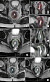
Figure 1.
Figure 1.
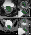
­Individual observer contours and consensus STAPLE contours for A-C) T2w imaging (axial, coronal, sagittal views), D-F) DWI (axial, coronal, sagittal views), and G-H) coregistered sessions (axial views T2w and DWI). Contour colours: red – T2w STAPLE; green – DWI STAPLE; cyan – CoReg STAPLE;dark blue, yellow, orange, pink – individual contours of observers 1-4.
Figure 2.
Figure 2.
­ Summary boxplot of individual observer and consensus (STAPLE) contour volumes across contouring sessions. Rad1: radiologist 1; Rad2: radiologist 2; RO1: radiation oncologist 1; RO2: radiation oncologist 2.
Figure 3.
Figure 3.
­Boxplot and summary statistics of observer-STAPLE comparisons for A) DSC, B) JI, C) HDmax, and D) MDA across delineation sessions.
Figure 4.
Figure 4.
­Scatterplot of generalised conformity index (CIgen) versus volume of delineations. Volume of the consensus STAPLE contour for each delineation is shown. CIgen significantly correlated with contour volume for T2w (r = 0.575, R2 = 0.330, p = 0.008) and CoReg GTV delineations (r = 0.532, R2 = 0.283, p = 0.016), but not for DWI or CoRegGTVs or subvolumes (SubV) (p>0.05).
Figure 5.
Figure 5.
­Outlier case due to peri-rectal collections with restricted diffusion on A) T2w, B) ADC and C) b800 image sequences. This was misattributed as extensive tumour spread by two observers on the DWI contouring session, by one observer on CoReg contouring, and not by any observer on T2w. Red: T2w STAPLE contour; Green: DWI STAPLE contour; Cyan: CoReg STAPLE contour.
Figure 6.
Figure 6.
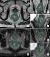
­Examplesof semi-automatically generated contours based on ADC threshold of 40thcentile, compared to manually-defined consensus STAPLE subvolumes.A-C) Axial,coronal and sagittal view of T2w MRI; D-F) axial, coronal and sagittal views ofADC. Cyan: whole tumour GTV (CoReg); purple: consensus (STAPLE) SubVdelineations; shaded green: thresholded volume.
Figure 7.
Figure 7.
­Examples of semi-automatically generated contours based on ADC threshold of 40th centile, compared to manually-defined consensus STAPLE subvolumes.A-C) Axial, coronal and sagittal view of T2w MRI; D-F) axial, coronal and sagittal views of ADC. Cyan: whole tumour GTV (CoReg); purple: consensus (STAPLE) SubV delineations; shaded green: thresholded volume
Figure 8.
Figure 8.
­Example of poor agreement between consensus STAPLE subvolume (SubV) delineation and ADC thresholding technique. Slight misregistration and changes in bowel gas between T2w and DWI scans result in inclusion of bowel lumen anteriorly as boost volume.Cyan: whole tumour GTV (CoReg); purple: consensus (STAPLE) SubV delineations; shaded green: 40th centile ADC thresholded volume.
Figure 9.
Figure 9.
­A-C) T2w and D-F) ADCimages of a favourable case with dedicated planning MRI. Images acquired co-axially with night-before bowel preparation.Manually-delineated and semi-automated delineation based on 40th centile ADC demonstrated substantial agreement (DSC: 0.801, JI: 0.668, HD: 7.12mm, MDA: 1.03mm). Cyan: whole tumour GTV (CoReg); purple: consensus (STAPLE) SubV delineations; shaded green: thresholded volumes

Similar articles

Cited by

References

    1. Burbach JPM, den Harder AM, Intven M, van Vulpen M, Verkooijen HM, Reerink O. Impact of radiotherapy boost on pathological complete response in patients with locally advanced rectal cancer: a systematic review and meta-analysis. Radiother Oncol 2014; 113: 1–9. doi: 10.1016/j.radonc.2014.08.035 - DOI - PubMed
    1. Hearn N, Atwell D, Cahill K, Elks J, Vignarajah D, Lagopoulos J, et al. . Neoadjuvant radiotherapy dose escalation in locally advanced rectal cancer: a systematic review and meta-analysis of modern treatment approaches and outcomes. Clin Oncol 2020;12 Jul 2020. doi: 10.1016/j.clon.2020.06.008 - DOI - PubMed
    1. Valentini V, Gambacorta MA, Cellini F, Aristei C, Coco C, Barbaro B, et al. . The INTERACT Trial: Long-term results of a randomised trial on preoperative capecitabine-based radiochemotherapy intensified by concomitant boost or oxaliplatin, for cT2 (distal)-cT3 rectal cancer. Radiother Oncol 2019; 134: 110–8. doi: 10.1016/j.radonc.2018.11.023 - DOI - PubMed
    1. Appelt AL, Pløen J, Vogelius IR, Bentzen SM, Jakobsen A. Radiation dose-response model for locally advanced rectal cancer after preoperative chemoradiation therapy. Int J Radiat Oncol Biol Phys 2013; 85: 74–80. doi: 10.1016/j.ijrobp.2012.05.017 - DOI - PMC - PubMed
    1. Pollard JM, Wen Z, Sadagopan R, Wang J, Ibbott GS. The future of image-guided radiotherapy will be Mr guided. Br J Radiol 2017; 90: 20160667. doi: 10.1259/bjr.20160667 - DOI - PMC - PubMed

MeSH terms