. 2020 Oct;586(7829):429-433.
doi: 10.1038/s41586-020-2719-5. Epub 2020 Sep 2.

STING cyclic dinucleotide sensing originated in bacteria

Affiliations

STING cyclic dinucleotide sensing originated in bacteria

Benjamin R Morehouse et al. Nature. 2020 Oct.

Abstract

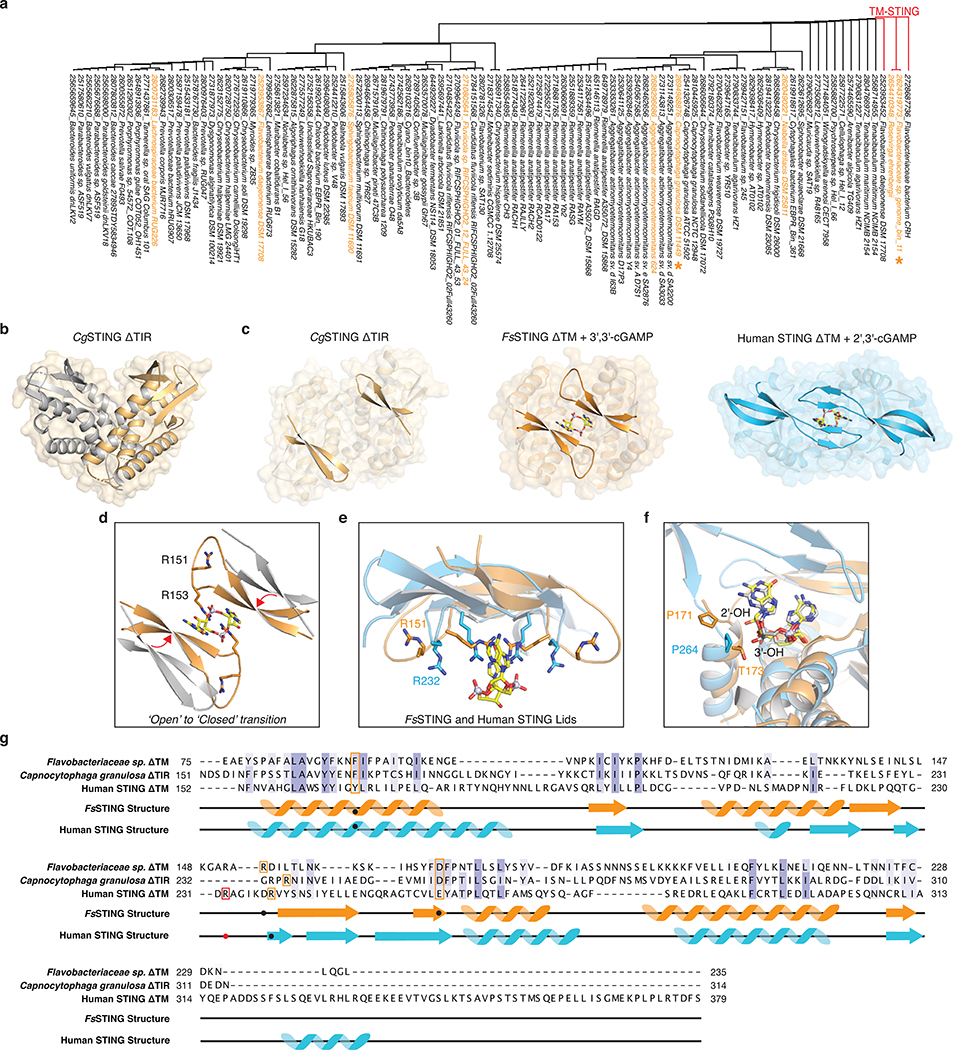
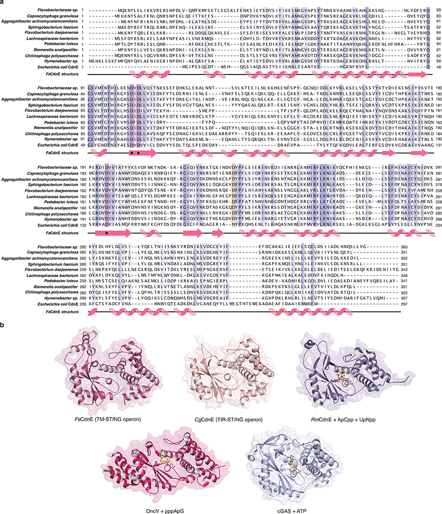
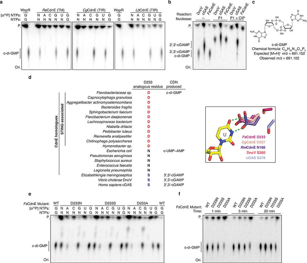
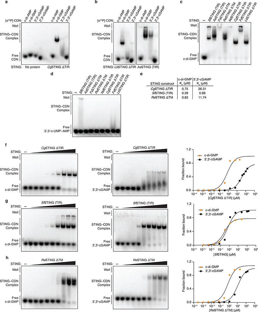
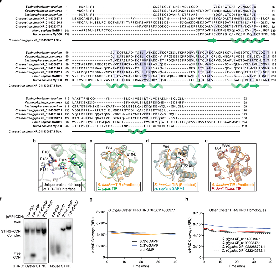
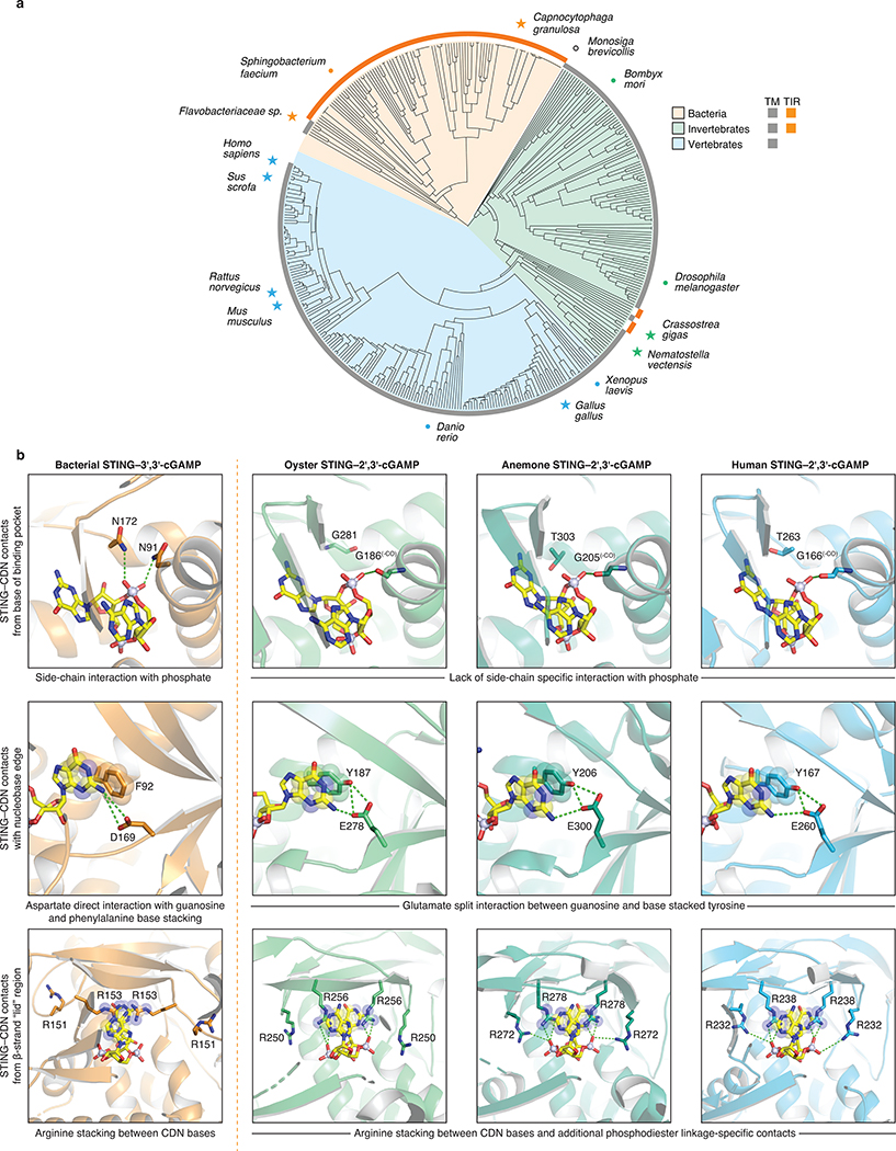
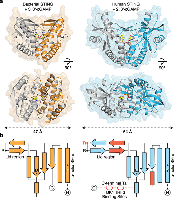
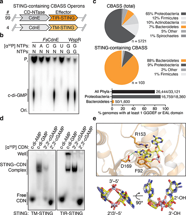
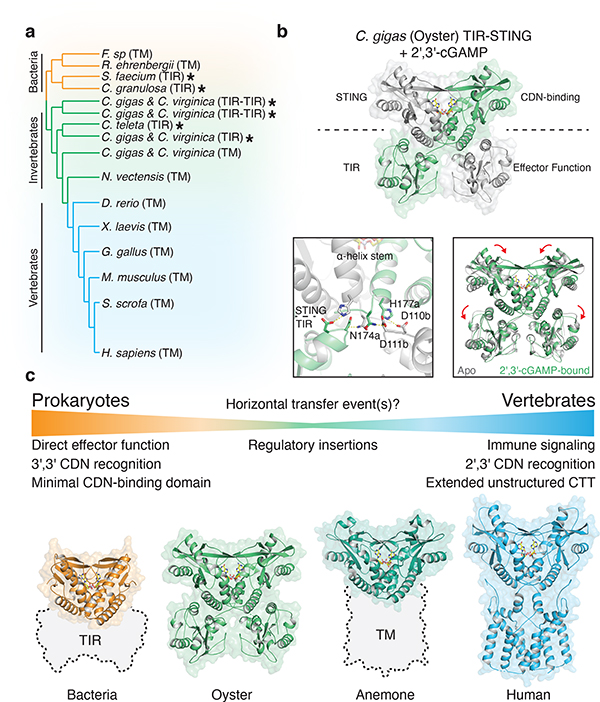
Stimulator of interferon genes (STING) is a receptor in human cells that senses foreign cyclic dinucleotides that are released during bacterial infection and in endogenous cyclic GMP-AMP signalling during viral infection and anti-tumour immunity1-5. STING shares no structural homology with other known signalling proteins6-9, which has limited attempts at functional analysis and prevented explanation of the origin of cyclic dinucleotide signalling in mammalian innate immunity. Here we reveal functional STING homologues encoded within prokaryotic defence islands, as well as a conserved mechanism of signal activation. Crystal structures of bacterial STING define a minimal homodimeric scaffold that selectively responds to cyclic di-GMP synthesized by a neighbouring cGAS/DncV-like nucleotidyltransferase (CD-NTase) enzyme. Bacterial STING domains couple the recognition of cyclic dinucleotides with the formation of protein filaments to drive oligomerization of TIR effector domains and rapid NAD+ cleavage. We reconstruct the evolutionary events that followed the acquisition of STING into metazoan innate immunity, and determine the structure of a full-length TIR-STING fusion from the Pacific oyster Crassostrea gigas. Comparative structural analysis demonstrates how metazoan-specific additions to the core STING scaffold enabled a switch from direct effector function to regulation of antiviral transcription. Together, our results explain the mechanism of STING-dependent signalling and reveal the conservation of a functional cGAS-STING pathway in prokaryotic defence against bacteriophages.


Conflict of interest statement

Author Information: The authors have no competing financial interests to declare. Correspondence and requests for materials should be addressed to P.J.K.

Figures

Extended Data Figure 1 |
Extended Data Figure 1 |. Structural analysis of bacterial STING–cyclic dinucleotide complex formation.
a, Phylogenetic tree of all CBASS-associated bacterial STING homologues based on structure-guided sequence alignment and previous bioinformatic analysis,. STING homologues investigated in this study are highlighted in orange, and a star denotes determined STING crystal structures. All TM-STING fusions cluster together. b, Crystal structure of a STING receptor from the bacterium Capnocytophaga granulosa (CgSTING) in the apo state reveals an open configuration with a solvent exposed cyclic dinucleotide-binding pocket at the dimeric interface (monomers in gold and grey for clarity). The CgSTING structure confirms that both divergent TM-STING and TIR-STING fusions are members of the same structurally conserved family of STING receptors. c, Comparison of the CgSTING, FsSTING–3′,3′-cGAMP, and human STING–2′,3′-cGAMP structures demonstrates conservation of an open-to-closed β-strand lid movement upon ligand binding. d, Overlay of the β-strand lid of CgSTING (grey) and FsSTING (orange) shows both inward translation and slight rotation resulting in a displacement of ~5 Å. R153 of FsSTING stacks between the bases of 3′,3′-cGAMP while R151 is splayed away from ligand. e, Comparison of the human STING and FsSTING lid region shows conserved contacts from β-strand arginine residues. Unlike in bacterial STING, human STING R232 makes an additional contact with the cyclic dinucleotide phosphodiester backbone that is critical for recognition of the 2′–5′ linkage in 2′,3′-cGAMP. See also Extended Data Figure 10b for detailed comparison. f, Modeling of 2′,3′-cGAMP into the FsSTING–3′,3′-cGAMP structure demonstrates an additional feature of bacterial STING preventing recognition of 2′–5′-linked cyclic dinucleotides. Although the overall cyclic dinucleotide conformation is shared between human and bacterial STING, the α-helix ending at P264 in human STING is a half turn longer in FsSTING (also ending in a proline) which places a conserved T173 residue in a position that occludes where the free 3′-OH of 2′,3′-cGAMP would be positioned. g, Structure-guided alignment of FsSTING and human STING cyclic dinucleotide binding domains. FsSTING and human STING exhibit no detectable sequence homology but share a conserved structural fold. Key residues involved in cyclic dinucleotide binding that are shared between bacterial and human STING are boxed in orange and human STING specific cyclic dinucleotide contacts are boxed in red. In FsSTING, D169 directly reads out the guanine base of c-di-GMP.
Extended Data Figure 2 |
Extended Data Figure 2 |. Structural analysis of STING-associated CD-NTase enzymes.
a, Sequence and secondary structure alignment of STING-associated CD-NTases reveals the extent of homology between CdnE homologues from unrelated bacterial strains. Highlighted positions include active-site residues (pink box), and an aspartic acid substitution at a position known to be involved in nucleotide substrate selection (orange box) that is unique to CD-NTases in STING-containing CBASS operons. A divergent E. coli CdnE that synthesizes cyclic UMP–AMP is included for comparison. b, Crystal structures of FsCdnE and CgCdnE from STING-containing CBASS operons allow direct comparison with previously determined bacterial and human CD-NTase structures. The FsCdnE and CgCdnE structures are most closely related to the Clade E CD-NTase structure from Rhodothermus marinus CdnE (RmCdnE). RmCdnE PDB 6E0L, V. cholerae DncV PDB 4TY0, human cGAS PDB 6CTA (DNA omitted for clarity).
Extended Data Figure 3 |
Extended Data Figure 3 |. Biochemical analysis of c-di-GMP synthesis by bacterial STING-associated CD-NTases.
a, In addition to FsCdnE (see Figure 2b), CdnE homologues from three divergent STING-containing CBASS operons were purified and tested for cyclic dinucleotide synthesis specificity using α32P-radiolabeled NTPs and thin-layer chromatography. Deconvolution experiments show a single major product requiring only GTP which migrates identically to c-di-GMP synthesized by the GGDEF enzyme WspR. All reactions were treated with alkaline phosphatase to remove exposed phosphates. Of note, only two bacterial genomes encoding a STING-containing CBASS operon retain proteins with a predicted canonical GGDEF c-di-GMP signaling domain. The exceptions are Chlorobi bacterium EBPR_Bin_190 which contains a single GGDEF domain that is fused to a SLATT domain and may be part of a CBASS-like system and a Lachnospiraceae bacterium RUG226 genome that encodes many GGDEF genes. The Lachnospiraceae bacterium RUG226 CdnE retains exclusive production of c-di-GMP suggesting the CdnE-STING system is sequestered in this bacterium or that an unknown mechanism may exist to prevent toxic STING activation. ReCdnE, Roseivirga ehrenbergii; CgCdnE, Capnocytophaga granulosa; LbCdnE, Lachnospiraceae bacterium; N, all four rNTPs; Pi, inorganic phosphate; Ori., origin. Data are representative of 2 independent experiments. b, Nuclease treatment confirms that the FsCdnE enzymatic product contains only canonical 3′–5′ phosphodiester bonds. The α32P-GTP cyclic dinucleotide product is susceptible to cleavage by nuclease P1 resulting in release of GMP as a new species which migrates further up the TLC plate. Further digestion with calf-intestinal phosphatase (CIP) removes all exposed phosphates resulting in complete loss of a labeled product spot. DncV (Dinucleotide cyclase in Vibrio) derived 3′,3′-cGAMP is similarly susceptible to complete digestion by P1 and CIP treatment whereas 2′,3′-cGAMP synthesized by mouse cGAS in only partially digested due to the presence of the non-canonical 2′–5′ bond. Data are representative of 2 independent experiments. c, High-resolution mass spectrometry analysis confirms the identity of the major FsCdnE enzymatic product as canonical c-di-GMP. Chemically synthesized c-di-GMP was used for direct spectral comparison. d, Sequence alignment and zoom-in inset of the active-site of the RmCdnE structure in complex with nonhydrolyzable UTP and ATP analogs (PDB 6E0L) demonstrating a contact in the CD-NTase lid domain known to control nucleobase sequence specificity. RmCdnE synthesizes cyclic UMP–AMP and uses N166 to specifically contact the uridine Watson-Crick edge. In contrast, FsCdnE and CgCdnE contain an aspartic acid substitution at this position and synthesize c-di-GMP, and Vibrio cholerae DncV and human cGAS contain a serine substitution at this position and synthesize 3′,3′-cGAMP and 2′,3′-cGAMP. 93% of STING-associated CD-NTase enzymes (96 of 103) contain a conserved aspartic acid at the FsCdnE D233 position consistent with strict specificity of c-di-GMP as the nucleotide second messenger controlling bacterial STING activation. RmCdnE PDB 6E0L, V. cholerae DncV PDB 4TY0, human cGAS PDB 6CTA (DNA omitted for clarity). e,f, Mutational analysis of the importance of D233 in FsCdnE c-di-GMP synthesis activity. D233 substitutions do not disrupt the overall ability of FsCdnE to selectively synthesis c-di-GMP, but a D233A substitution causes a mild reduction in nucleobase selectivity and efficiency of c-di-GMP synthesis. These results are consistent with a role for D233 in nucleobase selection but demonstrate full selectively is achieved by additional contacts in the active site pocket. Data are representative of 3 independent experiments.
Extended Data Figure 4 |
Extended Data Figure 4 |. Biochemical analysis of bacterial STING cyclic dinucleotide recognition specificity.
a,b, Electrophoretic mobility shift assay (EMSA) of purified bacterial STING proteins with radiolabeled cyclic dinucleotide (CDN) ligands. Bacterial STING receptors specifically recognize c-di-GMP and have a weak ability to bind 3′,3′-cGAMP. No interaction was observed with c-di-AMP or 2′,3′-cGAMP. Capnocytophaga granulosa (CgSTING), Lahnospiraceae bacterium STING (LbSTING), Aggregatibacter actinomyetemcomitans STING (AaSTING). Data are representative of 2 independent experiments. c, EMSA analysis of a diverse panel of bacterial STING homologues demonstrates conservation of c-di-GMP binding in both TM-STING and TIR-STING CBASS immunity. NdSTING (Niabella drilacis) and FdSTING (Flavobacterium daejeonense). Higher-order complex formation visible as well-shifted complexes is consistent with STING oligomerization results (see Figure 3f and Extended Data Figure 7). Data are representative of 3 independent experiments. d, EMSA analysis of diverse bacterial STING homologues broadly demonstrates no interaction with the 3′,3′-c-UMP–AMP second messenger synthesized by the divergent CD-NTase E. coli CdnE and further confirms the specificity of c-di-GMP signaling in bacterial STING-containing CBASS operons. Data are representative of 2 independent experiments. e,f,g,h, EMSA analysis and quantification of the affinity of bacterial STING homologues for c-di-GMP and 3′,3′-cGAMP. Signal intensity analysis is plotted as fraction bound (shifted / total signal) as a function of increasing protein concentration and fit to a single binding isotherm. CgSTING and ReSTING have a >10-fold preference for c-di-GMP while SfSTING has a similar apparent affinity for c-di-GMP and 3′,3′-cGAMP. Data are representative of 2 independent experiments.
Extended Data Figure 5 |
Extended Data Figure 5 |. Bacterial STING activation of TIR NADase activity.
a, HPLC analysis of chemical standards separated with an ammonium acetate:methanol gradient elution used to analyze bacterial TIR-STING activity (see Methods). The NAD+ and ADPr peaks have overlapping bases under these conditions. cADPr, cyclic adenosine diphosphate-ribose; ADPr, adenosine diphosphate-ribose; NAD+, β-nicotinamide adenine dinucleotide; NAM, nicotinamide. b, HPLC analysis of SfSTING NAD+ cleavage activity with gradient elution. SfSTING at 500 nM protein with 2 μM c-di-GMP converts 500 μM NAD+ into ADPr and NAM in 30 min at ambient temperature. SfSTING does not generate any cyclized product and is therefore a standard glycosyl hydrolase. Inset: Schematic of NAD+ cleavage reaction. c, HPLC analysis of chemical standards separated with an alternative isocratic elution strategy (see Methods) that results in clearer separation of NAD+ and ADPr peaks. d, HPLC analysis of SfSTING NAD+ cleavage activity with isocratic elution. SfSTING NAD+ cleavage activity requires specific activation with c-di-GMP (30 min reactions). e, HPLC analysis of SfSTING NAD+ cleavage activity and cyclic dinucleotide agonist specificity. Each reaction was tested with 500 nM SfSTING, 500 μM cyclic dinucleotide, and 500 μM NAD+ and sampled at 45, 90 or 180 min (gradient coloring in bars). SfSTING preferentially responds to c-di-GMP, but 3′,3′-cGAMP and c-di-AMP can function as weak agonists. Data are representative of 3 independent experiments. f, HPLC analysis of SfSTING NAD+ cleavage activity in the presence of 3′,3′-c-UMP–AMP. 3′,3′-c-UMP–AMP is a >1000× weaker agonist than c-di-GMP. Data are representative of 3 independent experiments. g, HPLC analysis of SfSTING NAD+ cleavage activity in the presence of a synthetic c-di-GMP analog with a noncanonical 2′–5′ linkage (2′,3′-c-di-GMP). 2′,3′-c-di-GMP is not capable of stimulating robust SfSTING activation even at very high concentrations (250 μM vs. 250 nM canonical c-di-GMP), confirming the specificity of bacterial STING for 3′–5′-linked cyclic dinucleotides. Data are representative of 3 independent experiments. h, Plate reader analysis of SfSTING NAD+ cleavage activity using the fluorescent substrate ε-NAD. ε-NAD increases in fluorescence intensity after cleavage. SfSTING exhibits rapid catalysis with complete turnover at 500 nM protein with 500 nM c-di-GMP after 10 min at 25°C. No background activity is observed in the absence of ligand. Data are representative of 3 independent experiments. i, Plate reader analysis of SfSTING NAD+ cleavage activity in the presence of 500 nM protein with increasing c-di-GMP concentration reveals that low nM c-di-GMP levels are sufficient to induce ε-NAD cleavage. Greater c-di-GMP concentrations are required for maximal activity consistent with binding data and the higher amount of c-di-GMP required for complete stabilization of the SfSTING–c-di-GMP complex (See Extended Data Figure 4e–h). Saturation occurs above 100 nM c-di-GMP with 40 min reactions. Data are ± s.d. of n = 3 technical replicates and are representative of 3 independent experiments. j,k, TIR-domain NAD+ cleavage activity requires protein oligomerization. For other systems,, TIR activation has been observed at very high in vitro protein concentrations or in the presence of affinity resins as an artificial oligomerization-inducing matrix,. We used a GST-TIR construct to express the SfSTING TIR domain in absence of the STING cyclic dinucleotide binding domain and observed that even at >200× the concentrations for which the full-length protein shows c-di-GMP induced activity, or in the presence of multivalent affinity resin, no NAD+ cleavage activity occurs. These results demonstrate NAD+ cleavage activity specifically requires STING cyclic dinucleotide recognition for activation. Data are representative of 2 independent experiments.
Extended Data Figure 6 |
Extended Data Figure 6 |. Mutagenesis analysis of bacterial STING cyclic dinucleotide recognition and TIR activation.
a, Table of bacterial STING cyclic dinucleotide contacts tested with mutagenesis analysis. Residues were selected according to contacts observed in the structure of the FsSTING–3′,3′-cGAMP complex (FsSTING residues listed in parentheses) and tested in SfSTING to allow analysis of the impact on both c-di-GMP binding and NADase activity. b, Plate reader analysis of mutant SfSTING NAD+ cleavage activity in the presence of 500 nM protein and increasing c-di-GMP concentration. Mutant SfSTING variants with cyclic dinucleotide binding pocket mutations require 10–1000× greater c-di-GMP concentration for NADase activation. Data are ± s.d. of n = 3 technical replicates and are representative of 3 independent experiments. c,d,e, EMSA analysis demonstrating that SfSTING R234A and D259A mutations reduce stable c-di-GMP complex formation compared to SfSTING WT (see Figure 2d). SfSTING D259A protein titration and quantification confirms significantly reduced affinity for c-di-GMP. Data are representative of 3 independent experiments. f,g, HPLC and plate reader analysis of mutant SfSTING NAD+ cleavage activity in the presence of 500 nM protein and 10 μM c-di-GMP (HPLC) or 500 nM protein and ± 20 μM c-di-GMP (plate reader). Mutation of residues responsible for ligand recognition in the cyclic dinucleotide binding domain (R234 and D259) and catalysis in the TIR enzymatic domain (E84) disrupts SfSTING NADase activity and explains loss of E. coli toxicity observed in Figure 3d. Note, 100 μM of c-di-GMP was used for SfSTING D259A plate reader NAD+ cleavage analysis to confirm complete loss of c-di-GMP-induced activation. HPLC data are representative of 3 independent experiments. Plate reader data are ± s.d. of n = 3 technical replicates and are representative of 3 independent experiments. h,l, Analysis of SfSTING toxicity in E. coli cells expressing normal c-di-GMP signaling enzymes with and without nicotinamide (NAM) supplementation. NAM supplementation is sufficient to partially alleviate SfSTING WT-induced NADase toxicity. Each line represents the average of 2 technical replicates for each of four separately outgrown colonies. Data are representative of 2 independent experiments.
Extended Data Figure 7 |
Extended Data Figure 7 |. Conservation of oligomerization as a mechanism of STING activation.
ai, STING SEC-MALS analysis. a,b, Full-length SfSTING changes oligomeric state in the presence of c-di-GMP or the weak agonist 3′,3′-cGAMP, and does not change oligomeric state in the presence of 2′,3′-cGAMP. c, With the TIR domain removed (ΔTIR), SfSTING no longer forms higher order complexes but notably remains dimeric in the apoprotein form. Bacterial TIR-STING proteins therefore appear to require the TIR domain to maintain stable higher-order oligomerization suggesting that intermolecular contacts are made with both TIR and STING domains. d, A TIR-only construct of SfSTING with the cyclic dinucleotide binding domain removed (ΔCDN) elutes as a single species which is consistent with the molecular weight for a homodimer. h, human STING (ΔTM) is a dimer in solution with or without 2′,3′-cGAMP, confirming that TM contacts are required for oligomerization and filament formation,. Nearly all tested bacterial and metazoan STING constructs migrate as dimers in solution consistent with the cyclic dinucleotide binding domain forming a constitutive homodimeric complex for ligand recognition. Two exceptions include ReSTING (ΔTM) and FsSTING (ΔTM) which form a mixture of monomeric and dimeric states in the absence of ligand and dimers or tetramers in the presence of 3′,3′-cGAMP. These results indicate that alternative oligomerization events may be required for activation of bacterial TM-STING effector function. j, Negative stain electron microscopy 2D class averages for SfSTING (E84A mutant) alone or in the presence of cyclic dinucleotide ligands. Stable STING filament formation requires c-di-GMP. 2D class averages were derived from particles selected from 75 micrographs for each condition. k, Representative micrograph images reveal extensive filament formation of varying length and orientation in the presence of c-di-GMP. Apo, c-di-AMP, and 2′,3′-cGAMP micrographs lack filaments. Images are each representative of n = 75 micrographs for each condition. l, Particle counting analysis of micrograph images shows that c-di-GMP induces more filament formation than 3′,3′-cGAMP and stable filament formation does not occur in the presence of c-di-AMP or 2′,3′-cGAMP. Data are mean ± s.d. for quantification of n = 4 groups of 10 micrograph images each.
Extended Data Figure 8 |
Extended Data Figure 8 |. Filament formation is required for bacterial TIR-STING activation.
a, Model of bacterial STING oligomerization and identification of surfaces involved in c-di-GMP-mediated filament formation. Electron microscopy analysis of SfSTING in the presence of c-di-GMP reveals filament formation likely occurs through parallel stacking of the homodimeric cyclic dinucleotide binding domain (see Extended Data Figure 7). To construct a potential model of this interaction, we used the X-ray crystal structure of the human STING–2′,3′-cGAMP complex (PDB 4KSY) and the cryo-electron microscopy structure of the chicken STING tetramer (PDB 6NT8) (top) as guides to position the FsSTING–3′,3′-cGAMP complex structure into a tetrameric conformation. The resulting model predicts that oligomerization-mediating surfaces in SfSTING include T200–N204, L275–F284, and G306–A310. b, EMSA analysis of SfSTING variants indicates that mutations to the predicted oligomerization surfaces do not prevent c-di-GMP recognition. SfSTING mutants tested include R307E/A309R, L201R/D203R, and the loop L275–Q282 replaced with a short GlySer linker (GSGGS). Data are representative of 2 independent experiments. c, Electron microscopy analysis of SfSTING variants in the presence of c-di-GMP. Mutations to the SfSTING surfaces identified in the structural model prevent all observable cyclic dinucleotide-induced filament formation supporting their predicted role in mediating oligomerization and bacterial STING filament formation. Images are each representative of n = 5 micrographs for each condition. d, HPLC analysis of mutant SfSTING NAD+ cleavage activity in the presence of 500 nM protein and 250 μM c-di-GMP for 3 h. In the absence of cyclic dinucleotide-mediated oligomerization all SfSTING NADase activity is lost, confirming the requirement of filament formation in TIR domain activation. Data are representative of 3 independent experiments.
Extended Data Figure 9 |
Extended Data Figure 9 |. Structural analysis of metazoan TIR-STING homologs.
a, Structure-guided alignment of the TIR domain in oyster TIR-STING with reference bacterial and metazoan TIR-domain-containing proteins. SARM1 is an example of a human TIR domain that catalyzes NAD+ cleavage and MyD88 is an example of a human TIR domain that signals through protein–protein interaction. The catalytic glutamate responsible for supporting NAD+ cleavage is conserved at the same spatial position among bacterial and oyster TIRs but is mutated in MyD88 (green box). However, it is not currently possible to predict from structure or sequence alone if TIR domains have enzymatic activity. b, Distinct from other TIR domain structures, the TIR domain in oyster TIR-STING contains a proline-rich loop region at the interface suggesting a specific role in dimer stabilization. c, Superposition of a homology model of the SfSTING TIR-domain with the TIR-domain of oyster TIR-STING shows the predicted catalytic glutamates for both proteins occupy distinct locations in the TIR fold. d, Superposition of a homology model of the SfSTING TIR-domain compared to the crystal structure of human SARM1 bound to ribose implies that different NAD+ binding pockets may exist between bacterial and eukaryotic TIRs as previously suggested. e, Superposition of a homology model of the SfSTING TIR-domain with the bacterial TIR domain from P. dentrificans shows a high degree of similarity. No crystal structures are available for bacterial TIR domains in an active state preventing identification of a specific mechanism of catalytic activation. f, EMSA analysis of oyster TIR-STING and mouse STING demonstrates a wide preference for cyclic dinucleotide interactions and clear ability to recognize the mammalian cGAS product 2′,3′-cGAMP. Data are representative of 3 independent experiments. g,h, Oyster TIR-STING which binds all tested CDNs does not exhibit NAD+ cleavage activity even at 10× the protein and ligand concentrations used to achieve robust activity with bacterial TIR-STING. We tested four other oyster TIR-STING homologues and observed no cyclic dinucleotide-stimulated NAD+ cleavage activity. These results support a potential switch in TIR-dependent protein–protein interactions to control downstream signaling similar to the TIR domain in human MyD88. Data are representative of 2 independent experiments.
Extended Data Figure 10 |
Extended Data Figure 10 |. Structure-guided analysis of STING phylogenetic conservation and cyclic dinucleotide recognition.
a, Structure-guided alignment and phylogenetic tree of STING proteins across bacterial and metazoan kingdoms. Bacterial STING homologues form a distinct cluster separate from all metazoan STING sequences and are mostly represented by TIR-STING fusions. A single STING-domain containing protein was identified in the choanoflagellate Monosiga brevicollis (denoted as an open black circle as this species is outside of the kingdom metazoan), no STING-domain containing proteins were found in archaea. TIR-STING fusions are rare in eukaryotes and cluster among invertebrate metazoans. No TIR-STING examples occur in vertebrates. Specific species of interest are highlighted to show the breadth of sequence diversity and stars mark proteins with available structures. b, Direct comparison of bacterial, oyster, anemone, and human STING crystal structures reveals conservation of specific cyclic dinucleotide contacts and critical differences in phosphodiester linkage recognition. Stacking interactions formed with the cyclic dinucleotide nucleobase face, aromatic side chains at the top of the α-helix stem, and arginine residues extending downward from the lid region are major conserved features shared between bacterial and metazoan STING proteins. Nucleotide-specific contacts are divergent between distinct STING structures, but notably the critical D169 guanosine N2-specific contact present in bacterial STING is conserved with a glutamic acid side chain in nearly all metazoan STING proteins. A critical feature absent in bacterial STING receptors is additional arginine-specific contacts to the phosphodiester backbone. The human STING R232 side chain contact known to be critical for high-affinity interactions with 2′,3′-cGAMP is conserved throughout metazoan STINGs representing a unique adaptation not found within bacterial STING receptors. Bacterial STING–3′,3′-cGAMP (FsSTING), oyster STING–2′,3′-cGAMP (C. gigas), sea anemone STING–2′,3′-cGAMP (N. vectensis PDB 5CFQ), human STING–2′,3′-cGAMP (H. sapiens PDB 4KSY).
Figure 1 |
Figure 1 |. Structure of bacterial STING and definition of metazoan-specific insertions.
a, Crystal structure of a STING receptor from the bacterium Flavobacteriaceae sp. (orange) in complex with the cyclic dinucleotide 3′,3′-cGAMP. The FsSTING–3′,3′-cGAMP structure demonstrates a conserved mechanism of cyclic dinucleotide sensing shared between bacteria and human cells, and allows direct comparison with the human STING–2′,3′-cGAMP complex (PDB 4KSY, blue). For clarity, one monomer of each homodimer is depicted in grey. b, STING topology diagrams denoting α-helices (rectangles), β-strands (arrows), and residues important for ligand recognition (FsSTING F92, D169, R153; hSTING Y167, E260, R232 in red, R238). Bacterial STING receptors reveal a minimal protein architecture required for cyclic dinucleotide sensing and allow direct definition of the structural insertions (red) in metazoan STING sequences required for signaling in animal cells.
Figure 2 |
Figure 2 |. Bacterial STING systems signal through the 3′–5′-linked nucleotide second messenger c-di-GMP.
a, Schematic of STING-containing CBASS prokaryotic defense operons. STING is encoded downstream of a Clade E CD-NTase nucleotide second messenger synthase (CdnE) and exists as a fusion protein appended to Toll/Interleukin-1 receptor (TIR) or transmembrane (TM) effector modules with 99 and 4 sequences found, respectively. b, Thin-layer chromatography analysis of Flavobacteriaceae sp. CdnE (FsCdnE) nucleotide second messenger synthesis. CdnE enzymes in STING-containing CBASS operons require GTP for catalysis and specifically synthesize the 3′–5′-linked product c-di-GMP. Control c-di-GMP synthesis reactions performed with the GGDEF enzyme P. aeruginosa WspR. N, all four radiolabeled NTPs; Pi, inorganic phosphate; Ori., origin. Data are representative of 3 independent experiments. c, Analysis of the genomic context of all STING-containing CBASS operons. STING-containing CBASS operons in bacteria are occur mainly in Bacteroidetes (top). Sequenced Bacteroidetes genomes are generally devoid of canonical GGDEF and EAL c-di-GMP signaling domains (bottom) and 97 of 99 STING-containing bacteria lack any other predicted c-di-GMP signaling component. Loss of canonical c-di-GMP signaling provides an explanation for co-option of c-di-GMP as a CD-NTase immune signal. Numbers near bar graphs denote genomes containing at least one gene with a GGDEF or EAL domain, out of total genomes in the analyzed database. d, Electrophoretic mobility shift assay monitoring bacterial STING–cyclic dinucleotide complex formation. Bacterial TM-STING and TIR-STING proteins preferentially recognize c-di-GMP and exhibit weaker affinity for 3′,3′-cGAMP. Bacterial STING receptors are unable to recognize the mammalian second messenger 2′,3′-cGAMP. TM-STING (R. ehrenbergii, ΔTM) and TIR-STING (S. faecium, full-length). Data are representative of 3 independent experiments. e, Zoom-in cutaway of the cyclic dinucleotide binding pocket in the FsSTING–3′,3′-cGAMP structure. Top, FsSTING makes sequence-specific contacts to the guanine base consistent with preferential c-di-GMP recognition. Bottom, bacterial STING recognizes 3′,3′-cGAMP (yellow) in a compact conformation similar to 2′,3′-cGAMP (grey) in complex with human STING (PDB 4KSY).
Figure 3 |
Figure 3 |. Cyclic dinucleotide-recognition controls bacterial STING oligomerization and TIR NAD+ cleavage activity.
a, Analysis of S. faecium TIR-STING (SfSTING) NAD+ cleavage activity using the fluorescent substrate ε-NAD. TIR-STING activity is potently stimulated in the presence of c-di-GMP (0, 5, 10, 15, 20 nM, 20 μM). Data are mean ± s.e.m. for n = 3 biological replicates. b, HPLC analysis of TIR-STING NAD+ cleavage. SfSTING cleaves NAD+ into the products ADPr and NAM, and activity is strictly dependent on the TIR active-site residue E84. Data are representative of 3 independent experiments. c, Quantification of SfSTING activity and comparison with previously characterized NADase and glycosyl hydrolase enzymes human SARM1 (hSARM1), Mycobacterium tuberculosis TNT, and Streptococcus pyogenes SPN. Data are mean for n = 2 independent replicates and are representative of 3 independent experiments. d, Analysis of SfSTING toxicity in E. coli cells expressing normal c-di-GMP signaling enzymes. Expression of SfSTING from an arabinose-inducible promoter (induced) results in a potent growth arrest phenotype. Toxicity is lost with a D259A mutation to the SfSTING cyclic dinucleotide-binding domain that inhibits c-di-GMP recognition. Each line represents the average of 2 technical replicates for each of four separately outgrown colonies. Data are representative of 2 independent experiments. e, Negative stain electron microscopy (EM) analysis and quantification demonstrates that SfSTING forms stable filaments only in the presence of c-di-GMP. Data are mean ± s.d. for quantification of n = 4 groups of 10 micrograph images. f, Electron microscopy analysis of SfSTING filament formation. Top, 2D class averages demonstrate that SfSTING forms well-ordered filaments in the presence of c-di-GMP and smaller fragmented assemblies in the presence of 3′,3′-cGAMP. Bottom, SfSTING–c-di-GMP filaments can be >30 nm in length. 2D class averages were derived from particles selected from 75 micrographs for each condition.
Figure 4 |
Figure 4 |. Structural basis of STING adaptation for signaling regulation.
a, Schematic derived from a structure-guided alignment of all known bacterial and metazoan STING sequences. Black stars denote TIR-STING fusions encoded in bacterial and metazoan genomes. Oysters encode several TIR-STING variants including genes predicted to contain multiple TIR domains (TIR–TIR). b, Crystal structure of the full-length C. gigas (oyster) TIR-STING receptor (Genbank sequence XP_011430837.1) in complex with 2′,3′-cGAMP. Top, oyster TIR-STING adopts a domain-swapped conformation with TIR domains appended across a central dimeric axis. Bottom left, 2′,3′-cGAMP recognition results in new interactions between the STING domain α-helix stem of one monomer and the TIR domain of the other monomer. Bottom right, Ligand binding induces STING domain lid closure and a downward rotation that repositions the appended TIR domains. c, Comparative structural analysis of STING adaptation suggests a model for the origin of cyclic dinucleotide sensing in innate immunity. STING evolved as a c-di-GMP sensor in prokaryotic bacteriophage defense, and acquisition in early metazoan cells may have allowed recognition of bacterial cyclic dinucleotides. Metazoan-specific structural insertions adapted STING for recognition of endogenous 2′,3′-cGAMP signaling and enabled antiviral and anti-tumor immune signaling in vertebrate cells. Structures shown: bacterial STING–3′,3′-cGAMP (FsSTING), oyster STING–2′,3′-cGAMP (C. gigas), sea anemone STING–2′,3′-cGAMP (N. vectensis PDB 5CFQ), human STING–2′,3′-cGAMP (H. sapiens PDB 6NT5 modeled with 6NT7).

Comment in

  • Bacteria sting viral invaders.
    Jenson J, Chen ZJ. Jenson J, et al. Nature. 2020 Oct;586(7829):363-364. doi: 10.1038/d41586-020-02712-8. Nature. 2020. PMID: 32989308 No abstract available.

References

    1. Ishikawa H & Barber GN STING is an endoplasmic reticulum adaptor that facilitates innate immune signalling. Nature (2008) 455, 674–678. - PMC - PubMed
    1. Zhong B, Yang Y, Li S, Wang YY, Li Y, Diao F, Lei C, He X, Zhang L, Tien P & Shu HB The adaptor protein MITA links virus-sensing receptors to IRF3 transcription factor activation. Immunity (2008) 29, 538–550 - PubMed
    1. Burdette DL, Monroe KM, Sotelo-Troha K, Iwig JS, Eckert B, Hyodo M, Hayakawa Y & Vance RE STING is a direct innate immune sensor of cyclic di-GMP. Nature (2011) 478, 515–518. - PMC - PubMed
    1. Sun L, Wu J, Du F, Chen X & Chen ZJ Cyclic GMP-AMP synthase is a cytosolic DNA sensor that activates the type I interferon pathway. Science (2013) 339, 786–791. - PMC - PubMed
    1. Ablasser A & Chen ZJ cGAS in action: Expanding roles in immunity and inflammation. Science (2019) 363 - PubMed

Publication types