. 2020 Aug 31;9(9):1999.
doi: 10.3390/cells9091999.

Evaluation of Multi-Layered Pancreatic Islets and Adipose-Derived Stem Cell Sheets Transplanted on Various Sites for Diabetes Treatment

Affiliations

Evaluation of Multi-Layered Pancreatic Islets and Adipose-Derived Stem Cell Sheets Transplanted on Various Sites for Diabetes Treatment

Yu Na Lee et al. Cells. .

Abstract

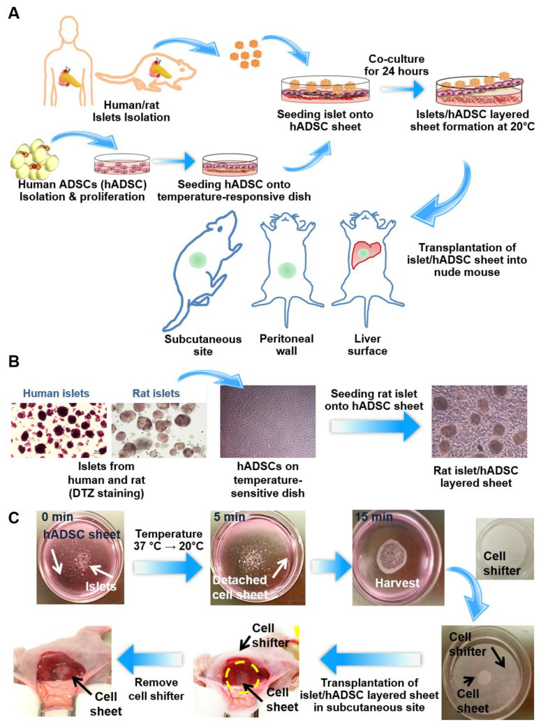
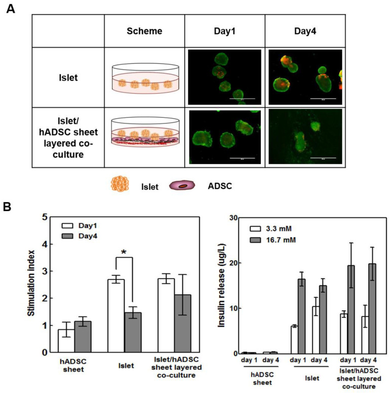
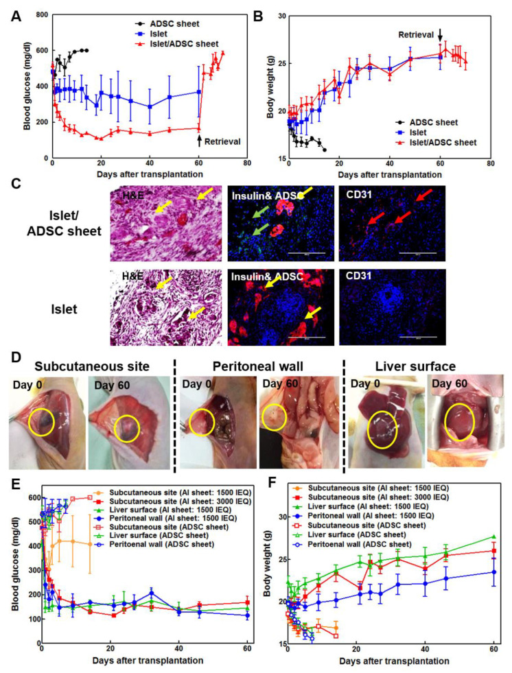
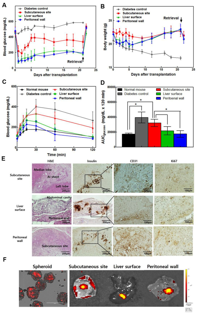
Islet cell transplantation is considered an ideal treatment for insulin-deficient diabetes, but implantation sites are limited and show low graft survival. Cell sheet technology and adipose-derived stem cells (ADSCs) can be useful tools for improving islet cell transplantation outcomes since both can increase implantation efficacy and graft survival. Herein, the optimal transplantation site in diabetic mice was investigated using islets and stem cell sheets. We constructed multi-layered cell sheets using rat/human islets and human ADSCs. Cell sheets were fabricated using temperature-responsive culture dishes. Islet/ADSC sheet (AI sheet) group showed higher viability and glucose-stimulated insulin secretion than islet-only group. Compared to islet transplantation alone, subcutaneous AI sheet transplantation showed better blood glucose control and CD31+ vascular traits. Because of the adhesive properties of cell sheets, AI sheets were easily applied on liver and peritoneal surfaces. Liver or peritoneal surface grafts showed better glucose control, weight gain, and intraperitoneal glucose tolerance test (IPGTT) profiles than subcutaneous site grafts using both rat and human islets. Stem cell sheets increased the therapeutic efficacy of islets in vivo because mesenchymal stem cells enhance islet function and induce neovascularization around transplanted islets. The liver and peritoneal surface can be used more effectively than the subcutaneous site in future clinical applications.

Keywords: adipose-derived stem cell; islet; liver surface; multi-layered cell sheet; peritoneal wall; subcutaneous site; transplantation.


Conflict of interest statement

Teruo Okano is a founder and director of the Scientific Advisory Board of CellSeed Inc., which has licenses for certain cell sheet-related technologies and patents from Tokyo Women’s Medical University. Yu Na Lee, Hye-Jin Yi, Yang Hee Kim, Song Lee, Jooyun Oh, In Kyong Shim, and Song Cheol Kim declare no potential conflict of interest. The authors declare that there are no other financial or non-financial competing interests. The authors of the article do not have any commercial association (e.g., consultancies, stock ownership, equity interests, or patent-licensing arrangements) that might pose a conflict of interest in connection with the submitted article.

Figures

Figure 1
Figure 1
Multi-layered islet/adipose-derived stem cell (ADSC) sheet co-culture model and transplantation in three different sites. (A) Schematic diagram of layered Islet/ADSC (AI) sheet co-culture. Islets were isolated from humans and rats. Human ADSCs (hADSCs) were isolated from human fat tissue and cultured on temperature-response dishes to produce cell sheets. Isolated islets were seeded on ADSC sheets after the hADSCs reached confluence and were harvested after layered co-culture for 24 h. AI sheets were transplanted in the subcutaneous site, peritoneal wall, and liver surface. (B) Microscope image of isolated islets, an hADSC sheet, and an AI multi-layered sheet. (C) Experimental procedure of layered AI sheet co-culture and transplantation into the subcutaneous site.
Figure 2
Figure 2
AI sheet groups showed better viability and glucose response than the islet-only group during the culture period. (A) The viability of islets and AI sheets was assessed by LIVE/DEAD staining. (B) Insulin release at 3.3 mM and 16.7 mM glucose in the hADSC sheet, islets, and AI sheet (in triplicate). The glucose-stimulated insulin secretion (GSIS) test showed better glucose response by the AI sheet group than by the islet-only group on days 1 and 4. Hundred hand-picked rat islets were used for each well. Results are presented as mean ± SD. Statistical significance was determined by a t-test of the stimulation index on days 1 and 4; * p < 0.05, n = 5.
Figure 3
Figure 3
(A) Subcutaneous transplantation of rat islets with ADSC sheets showed better blood glucose control than transplantation of islets alone in diabetic nude mice (n = 5). Rat islets (3000 IEQ) with ADSC sheet showed more favorable blood glucose levels than islets alone. (B) The body weight of the islet-only and AI sheet groups was increased compared to that of the ADSC control group. Islet and AI sheet group showed significantly lower blood glucose levels and higher body weight (p < 0.05). (C) Hematoxylin-eosin (H&E), insulin, and CD31 staining of tissues from mice transplanted with AI sheet and islets only. In the AI sheet group, ADSCs adequately surrounded the transplanted islets and induced angiogenesis (upper panel) compared to islet-only transplants (lower panel). Yellow arrow: islets, green arrow: ADSCs, red arrow: vessels. Scale bar: 200 µm. (D) Transplantation of AI sheet on the subcutaneous site (n = 5), peritoneal wall (n = 4), and liver surface (n = 5) was performed successfully. (E,F) Blood glucose levels and body weights after transplanting ADSC sheet on the subcutaneous site (3000 IEQ and 1500 IEQ), liver surface (1500 IEQ), and peritoneal wall (1500 IEQ). Mouse transplanted with ADSC sheet without islets at each transplantation sites are sham operation control (n = 3). Mouse transplanted with 1500 IEQ AI sheet showed high blood glucose level and weight loss, indicating that 1500 IEQ islet is not enough to control diabetes at subcutaneous site. However, 1500 IEQ AI sheet transplanted on liver surface or peritoneal wall could reduce blood glucose level to that of normal glycemia. The 3000 IEQ AI sheet also showed normal glycemia. Body weight of subcutaneous site (AI sheet: 3000 IEQ) and liver surface (AI sheet: 1500 IEQ) groups is statistically higher than that of subcutaneous site (AI sheet: 1500 IEQ), peritoneal wall (AI sheet: 1500 IEQ), and sham operation (ADSC sheet) groups. Blood glucose level of subcutaneous site (AI sheet: 3000 IEQ), liver surface (AI sheet: 1500 IEQ), and peritoneal wall (AI sheet: 1500 IEQ) groups is statistically higher than that of subcutaneous site (AI sheet: 1500 IEQ) and sham operation (ADSC sheet) groups. (p < 0.05).
Figure 4
Figure 4
The liver and peritoneal surface were more effective sites for cell sheet transplantation among subcutaneous, liver surface, and peritoneal wall sites using human islets (5000 IEQ) and ADSC multi-layered sheets in diabetic nude mice (n = 4). (A) Transplantation of AI sheets onto the three sites was performed successfully. The liver and peritoneal surface were more effective for controlling blood glucose levels. (B) The liver surface and peritoneal wall groups showed increased body weight. The subcutaneous group-maintained body weight, but diabetic control mice dramatically decreased in weight. (C) Intraperitoneal glucose tolerance test (IPGTT). The mice were fasted for 8 h, and then 2 g glucose/kg body weight was injected intraperitoneally. The baseline blood glucose was measured at 0, 15, 30, 60, and 120 min. The blood glucose profile (C) and area under the curve (AUC) (D) during IPGTT in the peritoneal wall were similar to those of non-diabetic mice. The liver surface and peritoneal wall groups showed significantly controllable results compared to the subcutaneous site group. Values are expressed as means ± SD. (* p < 0.05). (E) H&E and immunohistochemical staining for insulin, CD31, and Ki67 at 3 weeks after transplantation of AI sheets in three different sites. (F) PKH26 red fluorescent cell linker-labeled insulinoma spheroid with ADSC sheet was examined after transplantation with an in vivo imaging system.

References

    1. Ichii H., Inverardi L., Pileggi A., Molano R.D., Cabrera O., Caicedo A., Messinger S., Kuroda Y., Berggren P.-O., Ricordi C. A novel method for the assessment of cellular composition and beta-cell viability in human islet preparations. Am. J. Transplant. 2005;5:1635–1645. doi: 10.1111/j.1600-6143.2005.00913.x. - DOI - PubMed
    1. Alejandro R., Latif Z., Noel J., Shienvold F.L., Mintz D.H. Effect of anti-Ia antibodies, culture, and cyclosporin on prolongation of canine islet allograft survival. Diabetes. 1987;36:269–273. doi: 10.2337/diab.36.3.269. - DOI - PubMed
    1. Contreras J.L., Eckstein C., Smyth C.A., Bilbao G., Vilatoba M., Ringland S.E., Young C., Thompson J.A., Fernández J.A., Griffin J.H., et al. Activated protein C preserves functional islet mass after intraportal transplantation: A novel link between endothelial cell activation, thrombosis, inflammation, and islet cell death. Diabetes. 2004;53:2804–2814. doi: 10.2337/diabetes.53.11.2804. - DOI - PubMed
    1. Ryan E.A., Paty B.W., Senior P.A., Bigam D., Alfadhli E., Kneteman N.M., Lakey J.R.T., Shapiro A.M.J. Five-year follow-up after clinical islet transplantation. Diabetes. 2005;54:2060–2069. doi: 10.2337/diabetes.54.7.2060. - DOI - PubMed
    1. van der Windt D.J., Bottino R., Casu A., Campanile N., Cooper D.K.C. Rapid loss of intraportally transplanted islets: An overview of pathophysiology and preventive strategies. Xenotransplantation. 2007;14:288–297. doi: 10.1111/j.1399-3089.2007.00419.x. - DOI - PubMed

Publication types