. 2020 Aug 31;9(9):2001.
doi: 10.3390/cells9092001.

PIM1 Promotes Survival of Cardiomyocytes by Upregulating c-Kit Protein Expression

Affiliations

PIM1 Promotes Survival of Cardiomyocytes by Upregulating c-Kit Protein Expression

David E Ebeid et al. Cells. .

Abstract

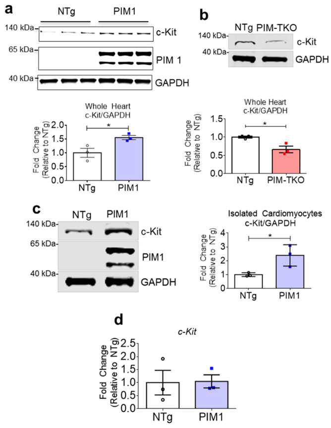
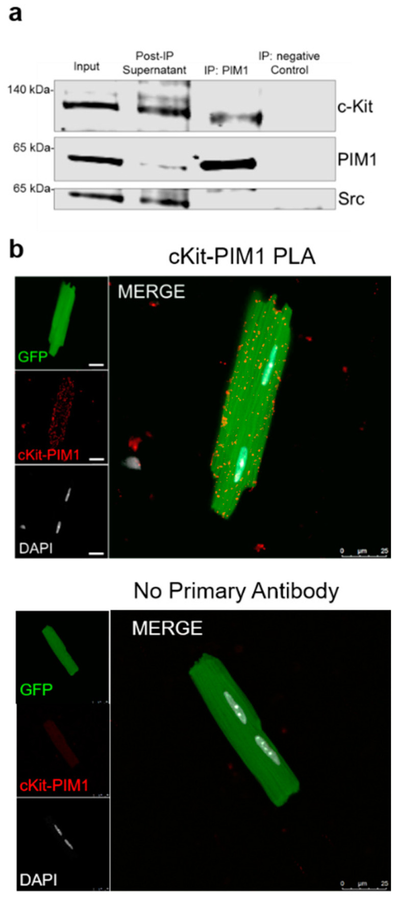
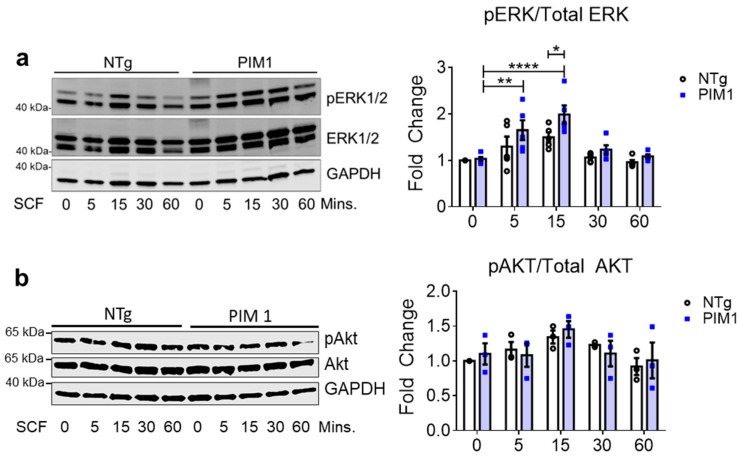
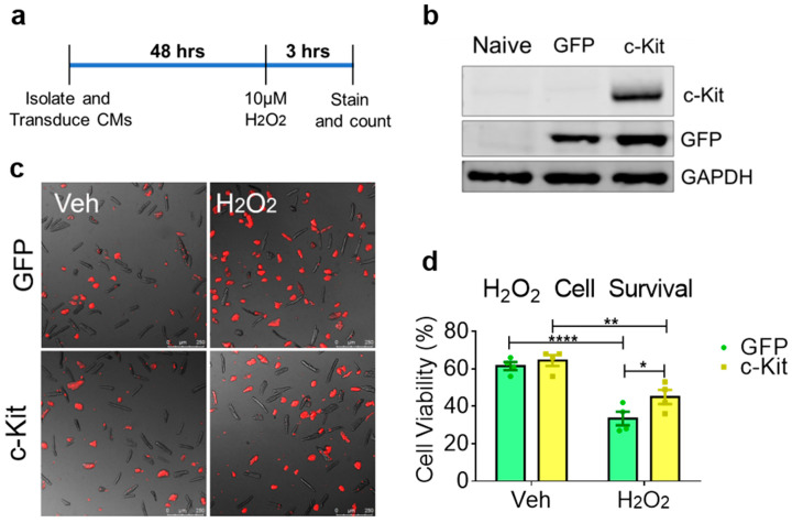
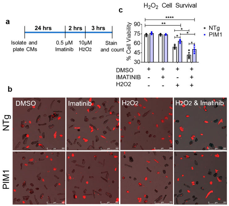
Enhancing cardiomyocyte survival is crucial to blunt deterioration of myocardial structure and function following pathological damage. PIM1 (Proviral Insertion site in Murine leukemia virus (PIM) kinase 1) is a cardioprotective serine threonine kinase that promotes cardiomyocyte survival and antagonizes senescence through multiple concurrent molecular signaling cascades. In hematopoietic stem cells, PIM1 interacts with the receptor tyrosine kinase c-Kit upstream of the ERK (Extracellular signal-Regulated Kinase) and Akt signaling pathways involved in cell proliferation and survival. The relationship between PIM1 and c-Kit activity has not been explored in the myocardial context. This study delineates the interaction between PIM1 and c-Kit leading to enhanced protection of cardiomyocytes from stress. Elevated c-Kit expression is induced in isolated cardiomyocytes from mice with cardiac-specific overexpression of PIM1. Co-immunoprecipitation and proximity ligation assay reveal protein-protein interaction between PIM1 and c-Kit. Following treatment with Stem Cell Factor, PIM1-overexpressing cardiomyocytes display elevated ERK activity consistent with c-Kit receptor activation. Functionally, elevated c-Kit expression confers enhanced protection against oxidative stress in vitro. This study identifies the mechanistic relationship between PIM1 and c-Kit in cardiomyocytes, demonstrating another facet of cardioprotection regulated by PIM1 kinase.

Keywords: PIM1; c-Kit; cardiomyocyte; cardioprotection.


Conflict of interest statement

The authors declare no conflict of interest.

Figures

Figure 1
Figure 1
PIM1 upregulates c-Kit protein expression. (a) Immunoblot analysis showing expression of c-Kit in PIM1 vs. NTg whole heart lysates with quantification below. N = 3, Error bars represent SEM, * p < 0.05 vs. untreated as measured by Student t test. (b) Immunoblot analysis showing the expression of c-Kit in PIM-TKO vs. NTg whole heart lysates with quantification shown below. N = 3, error bars represent SEM, * p < 0.05 vs. untreated as measured by Student t test. (c) Immunoblot analysis showing c-Kit expression in cardiomyocytes isolated from PIM1-overexpressing hearts vs. NTg with quantification on the right. N = 3, error bars represent SEM, * p < 0.05 vs. untreated as measured by Student t test. (d) Gene expression of c-Kit in cardiomyocytes isolated from PIM1 and NTg hearts as revealed by qPCR. N = 3, Error bars represent SEM.
Figure 2
Figure 2
PIM1 interacts with c-Kit. (a) Immunoblot analysis of a co-immunoprecipitation using whole heart lysates from PIM1 hearts in which PIM1 was pulled down. (b) Interaction between PIM1 and c-Kit as determined by proximity ligation assay (top panel) and in the absence of primary antibody (bottom panel). Proximity events are shown as red dots.
Figure 3
Figure 3
Cardioprotective signaling downstream of c-Kit is elevated in PIM1 cardiomyocytes. (a) Immunoblot analysis of activated ERK1/2 and (b) activated AKT in NTg and PIM1 cardiomyocytes following treatment with SCF over 60 min. Quantification is shown on the right. N = 5, Error bars represent SEM, * p < 0.05, ** p < 0.01 and **** p < 0.0001 as measured by two-way ANOVA multiple comparison with Tukey.
Figure 4
Figure 4
Cardiomyocytes with elevated c-Kit expression demonstrate enhanced resistance against oxidative stress. (a) Schematic showing the treatment procedure. (b) Immunoblot analysis of isolated cardiomyocytes transduced with GFP or c-Kit-GFP. (c) Fluorescent images of TO-PRO-3 staining on GFP or c-Kit-expressing cardiomyocytes treated with vehicle or H2O2 overlaid with brightfield images depicting dead or dying cells in red. (d) Quantification of cell viability counted from fluorescent images. N = 4, 300–600 cardiomyocytes counted per group, error bars represent SEM, * p < 0.05, ** p < 0.01 and **** p < 0.0001 as measured by two-way ANOVA multiple comparison with Tukey.
Figure 5
Figure 5
PIM1 and c-Kit act independently to confer cardioprotection. (a) schematic showing the treatment procedure. (b) Representative images of NTg and PIM1 cardiomyocytes stained with TO-PRO-3. Cells depicted in red are counted as non-viable cells. (c) Quantification of cell viability counted from fluorescent images. N = 5, 300–600 cardiomyocytes counted per group, error bars represent SEM, * p < 0.05, ** p < 0.01 and **** p < 0.0001 as measured by two-way ANOVA multiple comparison with Tukey.

References

    1. Li F., Wang X., Capasso J.M., Gerdes A.M. Rapid transition of cardiac myocytes from hyperplasia to hypertrophy during postnatal development. J. Mol. Cell. Cardiol. 1996;28:1737–1746. doi: 10.1006/jmcc.1996.0163. - DOI - PubMed
    1. Zebrowski D.C., Engel F.B. The cardiomyocyte cell cycle in hypertrophy, tissue homeostasis, and regeneration. Rev. Physiol. Biochem. Pharmacol. 2013;165:67–96. doi: 10.1007/112_2013_12. - DOI - PubMed
    1. Zebrowski D.C., Vergarajauregui S., Wu C.C., Piatkowski T., Becker R., Leone M., Hirth S., Ricciardi F., Falk N., Giessl A., et al. Developmental alterations in centrosome integrity contribute to the post-mitotic state of mammalian cardiomyocytes. Elife. 2015;4:e05563. doi: 10.7554/eLife.05563. - DOI - PMC - PubMed
    1. Kang M.J., Kim J.S., Chae S.W., Koh K.N., Koh G.Y. Cyclins and cyclin dependent kinases during cardiac development. Mol. Cells. 1997;7:360–366. doi: 10.1006/jmcc.1996.0163. - DOI - PubMed
    1. Poolman R.A., Gilchrist R., Brooks G. Cell cycle profiles and expressions of p21(CIP1) and p27(KIP1) during myocyte development. Int. J. Cardiol. 1998;67:133–142. doi: 10.1016/S0167-5273(98)00320-9. - DOI - PubMed

Publication types

Substances