. 2020 Oct 27;94(22):e00403-20.
doi: 10.1128/JVI.00403-20. Print 2020 Oct 27.

Retinoic Acid Inducible Gene I and Protein Kinase R, but Not Stress Granules, Mediate the Proinflammatory Response to Yellow Fever Virus

Affiliations

Retinoic Acid Inducible Gene I and Protein Kinase R, but Not Stress Granules, Mediate the Proinflammatory Response to Yellow Fever Virus

Guillaume Beauclair et al. J Virol. .

Abstract

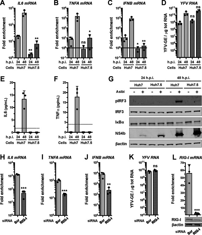
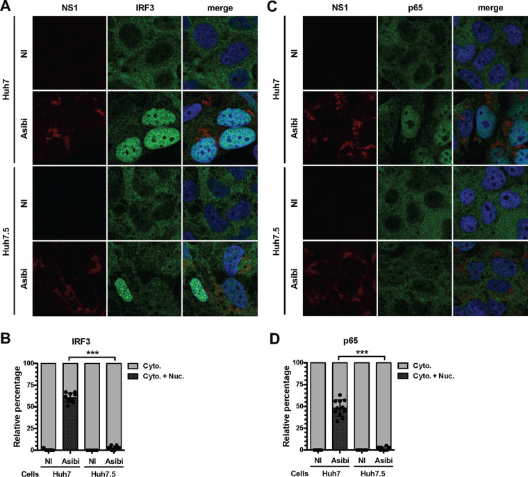
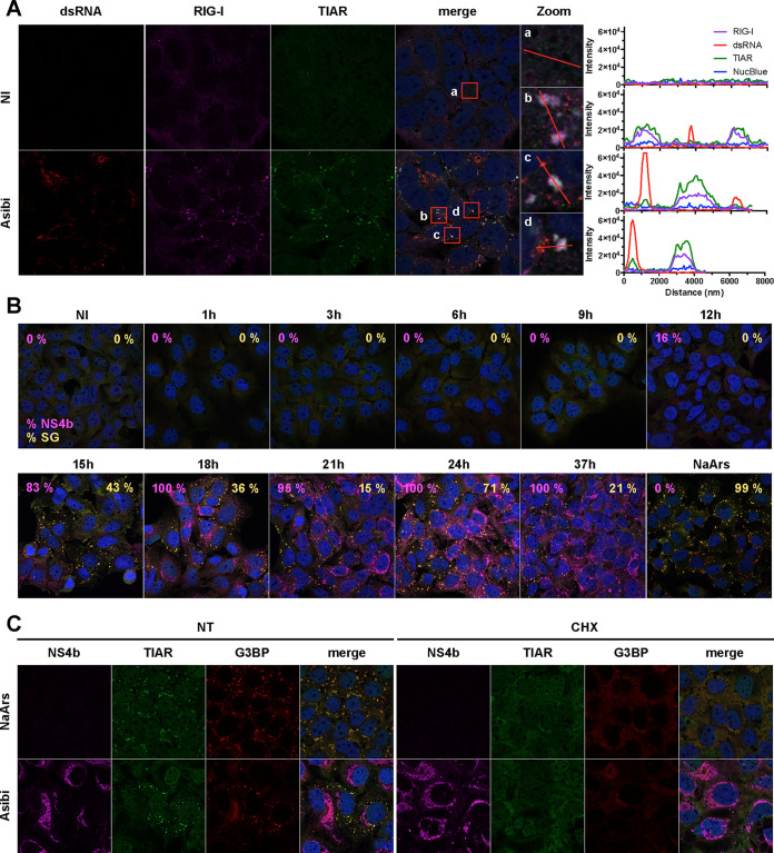
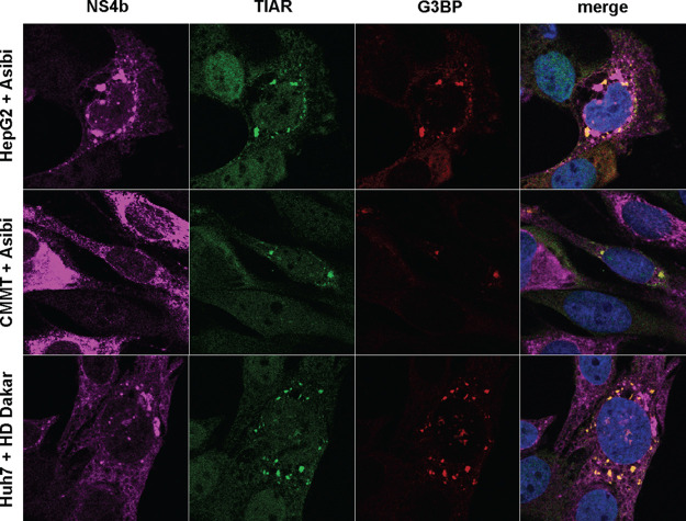
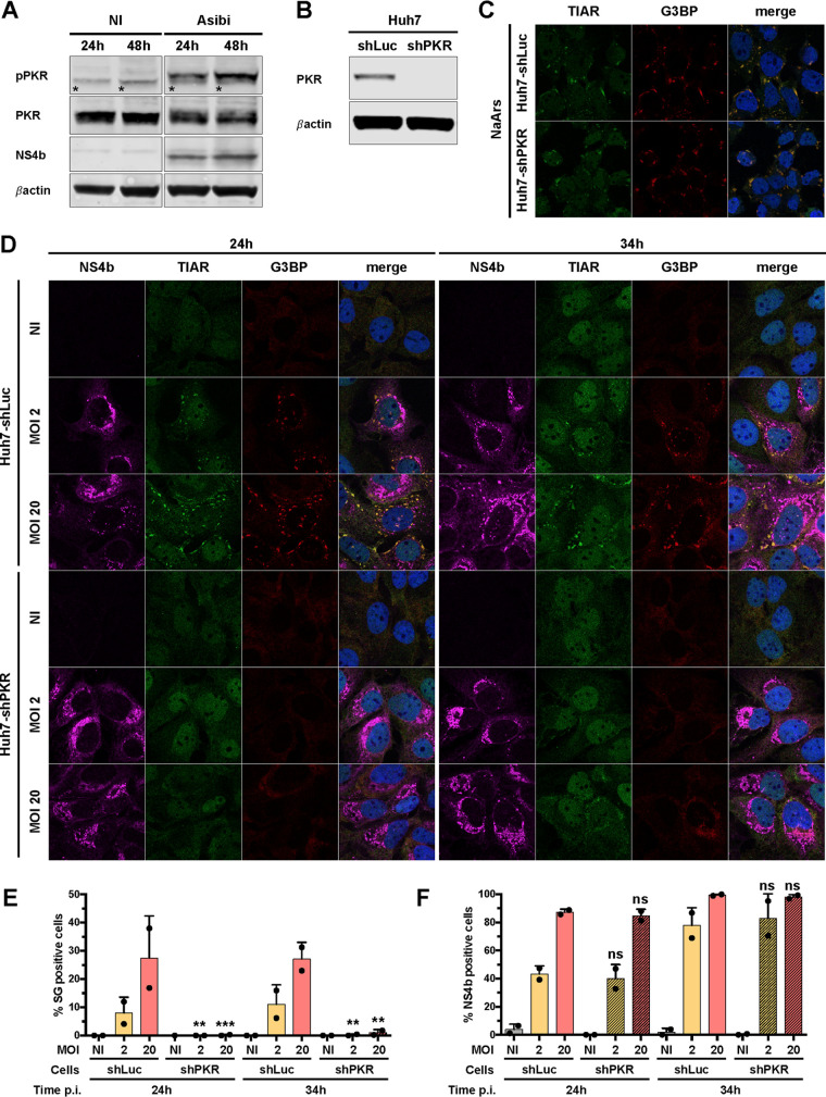
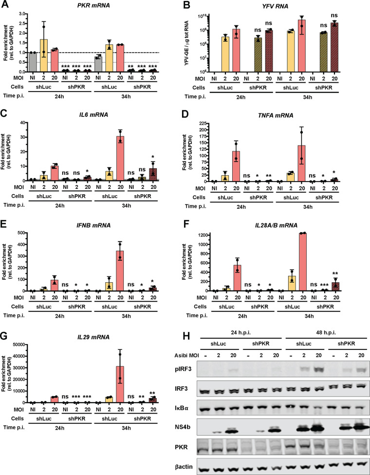
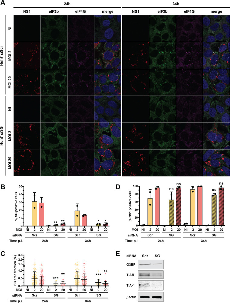
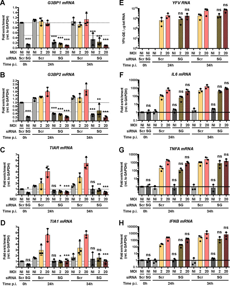
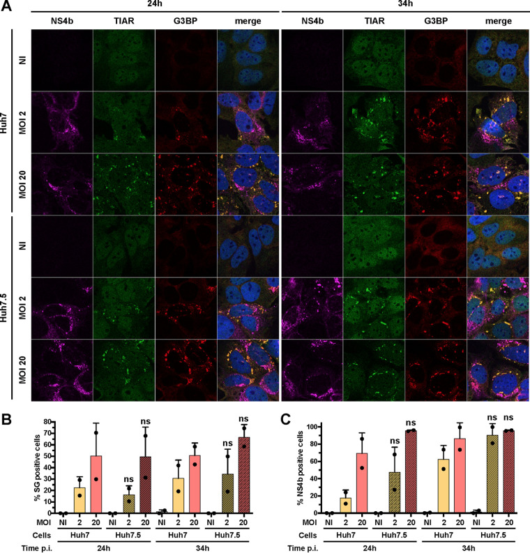
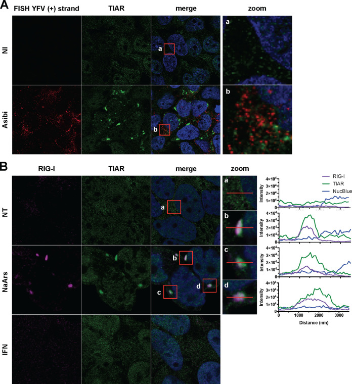
Yellow fever virus (YFV) is an RNA virus primarily targeting the liver. Severe YF cases are responsible for hemorrhagic fever, plausibly precipitated by excessive proinflammatory cytokine response. Pathogen recognition receptors (PRRs), such as the cytoplasmic retinoic acid inducible gene I (RIG-I)-like receptors (RLRs), and the viral RNA sensor protein kinase R (PKR), are known to initiate a proinflammatory response upon recognition of viral genomes. Here, we sought to reveal the main determinants responsible for the acute cytokine expression occurring in human hepatocytes following YFV infection. Using a RIG-I-defective human hepatoma cell line, we found that RIG-I largely contributes to cytokine secretion upon YFV infection. In infected RIG-I-proficient hepatoma cells, RIG-I was localized in stress granules. These granules are large aggregates of stalled translation preinitiation complexes known to concentrate RLRs and PKR and are so far recognized as hubs orchestrating RNA virus sensing. Stable knockdown of PKR in hepatoma cells revealed that PKR contributes to both stress granule formation and cytokine induction upon YFV infection. However, stress granule disruption did not affect the cytokine response to YFV infection, as assessed by small interfering RNA (siRNA)-knockdown-mediated inhibition of stress granule assembly. Finally, no viral RNA was detected in stress granules using a fluorescence in situ hybridization approach coupled with immunofluorescence. Our findings suggest that both RIG-I and PKR mediate proinflammatory cytokine induction in YFV-infected hepatocytes, in a stress granule-independent manner. Therefore, by showing the uncoupling of the cytokine response from the stress granule formation, our model challenges the current view in which stress granules are required for the mounting of the acute antiviral response.IMPORTANCE Yellow fever is a mosquito-borne acute hemorrhagic disease caused by yellow fever virus (YFV). The mechanisms responsible for its pathogenesis remain largely unknown, although increased inflammation has been linked to worsened outcome. YFV targets the liver, where it primarily infects hepatocytes. We found that two RNA-sensing proteins, RIG-I and PKR, participate in the induction of proinflammatory mediators in human hepatocytes infected with YFV. We show that YFV infection promotes the formation of cytoplasmic structures, termed stress granules, in a PKR- but not RIG-I-dependent manner. While stress granules were previously postulated to be essential platforms for immune activation, we found that they are not required for the production of proinflammatory mediators upon YFV infection. Collectively, our work uncovered molecular events triggered by the replication of YFV, which could prove instrumental in clarifying the pathogenesis of the disease, with possible repercussions for disease management.

Keywords: cytokines; flavivirus; innate immunity; interferons; liver inflammation; pattern recognition receptors; stress granules; yellow fever virus.


Figures

FIG 1
FIG 1
The Asibi strain of YFV triggers the production of proinflammatory cytokines in hepatoma cells through RIG-I. Huh7 and Huh7.5 cells were left uninfected or infected with Asibi at an MOI of 1 for 24 or 48 h. (A to C) The relative amounts of IL-6 (A), TNFA (B), and IFNB (C) mRNAs were determined by RT-qPCR analysis and normalized to GAPDH mRNA and noninfected samples. (D) The relative amounts of cell-associated viral RNA were determined by RT-qPCR analysis and expressed as genome equivalents (YFV-GE) per μg of total cellular RNA. (E and F) Culture media of the indicated cells were analyzed by ELISA to determine the amounts of secreted IL-6 (E) and TNF-α (F). The data are the means ± SD from two (Huh7.5) or three (Huh7) experiments. The dashed lines indicate the limits of detection of the IL-6 and TNF-α ELISAs (2 and 4 pg/ml, respectively). Statistical analysis compared Huh7.5 samples to Huh7 samples. (G) Whole-cell lysates of the indicated cells were analyzed by Western blotting with antibodies against the indicated proteins. (H to J) The relative amounts of IL-6 (H), TNFA (I), and IFNB (J) mRNAs were determined by RT-qPCR analysis in cells transfected with RIG-I-specific siRNAs or control siRNAs (Scr). The samples were normalized to GAPDH mRNAs. (K) The relative amounts of cell-associated viral RNA were determined by RT-qPCR analysis and expressed as genome equivalents (YFV-GE) per μg of total cellular RNA. (L) The relative amounts of RIG-I mRNAs were determined by RT-qPCR analysis in cells transfected with RIG-I-specific siRNAs or control siRNAs (Scr). The samples were normalized to GAPDH mRNAs. Bottom, the abundances of RIG-I and actin were determined by Western blotting in cells that were transfected with control or RIG-I-specific siRNAs. Whole-cell lysates were analyzed 36 h later. The RT-qPCR and ELISA data are the means ± SD from two or three independent experiments, as indicated by the dots on the graphs. ns, nonsignificant; *, P < 0.05; **, P < 0.01. Western blots are representative of two independent experiments.
FIG 2
FIG 2
RIG-I contributes to IRF3 and NF-κB activation in hepatoma cells infected with the Asibi strain of YFV. Huh7 or Huh7.5 cells were left uninfected (NI) or infected with YFV-Asibi for 48 h at an MOI of 20. Cells were then stained with antibodies recognizing NS1 (red), IRF3 (A and B), or p65 (C and D) (green), as well as with NucBlue (blue). Ten field containing around 30 cells were scored for nuclear translocation of IRF3 or p65. ns, nonsignificant; ***, P < 0.001.
FIG 3
FIG 3
RIG-I localizes to stress granules in Huh7 cells infected with YFV. (A) Huh7 cells were left uninfected (NI) or infected with Asibi at an MOI of 20 for 24 h. Cells were then stained with dsRNA (red), RIG-I (purple), or TIAR (green) antibodies and NucBlue (blue). (B) Huh7 cells were left uninfected (NI), treated with 0.5 mM NaArs for 30 min, or infected with Asibi at an MOI of 20 for the indicated time. Cells were then stained with NS4b (purple), G3BP (red), or TIAR (green) antibodies and NucBlue (blue). Percentages of NS4b-positive and G3BP-positive cells were estimated and are shown in purple and yellow, respectively. (C) Huh7 cells were treated with 0.5 mM NaArs for 30 min or infected with YFV-Asibi at an MOI of 20 for 48 h. Cells were then left untreated (NT) or treated with 100 μg/ml cycloheximide (CHX) for 1 h and stained with antibodies against NS4b (purple), TIAR (green), and G3BP (red), as well as with NucBlue (blue).
FIG 4
FIG 4
YFV infection induces the formation of stress granules in several mammalian cells. HepG2 or CMMT cells were infected with Asibi at an MOI of 20 for, respectively, 48 and 24 h. Huh7 cells were infected with YFV-HD Dakar at an MOI of 10 for 24 h. Cells were then stained with NS4b (purple), TIAR (green), or G3BP (red) antibodies and NucBlue (blue).
FIG 5
FIG 5
PKR contributes to stress granule formation in Asibi-infected Huh7 cells. (A) Huh7 cells were left uninfected (NI) or infected with Asibi at an MOI of 20 for 24 or 48 h. Whole-cell lysates were analyzed by Western blotting with antibodies against the indicated proteins. Samples were loaded on the same gel. *, unspecific bands. (B) Whole-cell lysates of Huh7-shLuc and Huh7-shPKR cells were analyzed by Western blotting with antibodies against the indicated proteins. (C) Huh7-shLuc and Huh7-shPKR cells were treated with 0.5 mM NaArs for 30 min and stained with TIAR (green) and G3BP (red) antibodies, as well as with NucBlue (blue). (D) Huh7 cells were left uninfected (NI) or infected with YFV-Asibi at an MOI of 2 or 20 for 24 or 34 h. Cells were then stained with NS4b (purple), TIAR (green), or G3BP (red) antibodies and NucBlue (blue). Images are representative of two independent experiments. (E and F) Percentages of G3BP-positive (E) or NS4b-positive (F) cells were estimated by analyzing at least 200 cells per condition. Statistical analysis compared Huh7-shPKR cells to Huh7-shLuc cells. ns, nonsignificant; *, P < 0.05; **, P < 0.01; ***, P < 0.001.
FIG 6
FIG 6
PKR contributes to innate response in Asibi-infected Huh7 cells. Huh7-shLuc or -shPKR cells were left uninfected (NI) or infected with Asibi at an MOI of 2 or 20 for 24 or 34 h. (A and C to G) The relative amounts of PKR (A), IL-6 (C), TNFA (D), IFNB (E), IL28A/B (F), and IL-29 (G) mRNAs were determined by qPCR analysis and normalized to that of GAPDH mRNA and noninfected shLuc samples. (B) The relative amounts of cell-associated viral RNA were determined by RT-qPCR analysis and expressed as genome equivalents (YFV-GE) per μg of total cellular RNA. Means ± SD are shown. One-way ANOVA on log-transformed data with Tukey-corrected multiple-comparison tests were performed. shPKR samples were compared to shLuc samples. ns, nonsignificant; *, P < 0.05; **, P < 0.01; ***, P < 0.001. (H) Whole-cell lysates of the indicated cells were analyzed by Western blotting with antibodies against the indicated proteins. The Western blot is representative of two independent experiments.
FIG 7
FIG 7
Knockdown of G3BP1, G3BP2, TIAR, and TIA1 inhibits stress granule formation. (A) Huh7 cells were transfected with scramble siRNA (siScr) or with siRNA targeting G3BP1, G3BP2, TIAR, and TIA1 (siSG) at 48 and 24 h prior to Asibi infection. Transfected cells were left uninfected (NI), treated with 0.5 mM NaArs for 30 min (NaArs), or infected with Asibi at an MOI of 2 or 20 for 24 or 34 h. Cells were then stained with NS1 (purple), eIF3b (green), or eIF4G (red) antibodies and NucBlue (blue). Images are representative of two independent confocal microscopy analyses. (B to D) Percentages of eIF4G- and eIF3b-positive (B) or NS1-positive (D) cells were estimated, and the stress granule area was determined for each cell as the sum of the whole area occupied by stress granules divided by the total cell surface (C). Around 200 cells per independent experiment were scored. Statistical analysis compared Huh7-siSG cells to Huh7-siScr cells. ns, nonsignificant; *, P < 0.05; **, P < 0.01; ***, P < 0.001. (E) The abundances of G3BP1/2, TIAR, TIA1, and actin were determined by Western blotting in Huh7 cells, which were transfected with control siRNAs or with siSG. Whole-cell lysates were analyzed with antibodies against the indicated proteins. Western blots are representative of two independent experiments.
FIG 8
FIG 8
Inhibition of stress granule formation has a limited effect on the antiviral response. Huh7 cells were transfected with scramble siRNA (siScr) or with four pools of siRNA targeting G3BP1, G3BP2, TIAR, and TIA1 (siSG) at 48 and 24 h prior to infection. Transfected cells were left uninfected (NI) or infected with Asibi at an MOI of 2 or 20. Cells were harvested prior to infection (0 h) and at 24 and 34 h postinfection. (A to D and F to H) The relative amounts of G3BP1 (A), G3BP2 (B), TIAR (C), TIA1 (D), IL-6 (F), TNFA (G), and IFNB (H) mRNAs were determined by qPCR analysis and normalized to that of GAPDH mRNA and noninfected shScr samples. (E) The relative amounts of cell-associated viral RNA were determined by RT-qPCR analysis and expressed as genome equivalents (YFV-GE) per μg of total cellular RNA. Means ± SD are shown. One-way ANOVA on log-transformed data with Tukey-corrected multiple-comparison tests were performed. Samples treated with siSG RNA were compared to siScr samples. ns, nonsignificant; *, P < 0.05; **, P < 0.01; ***, P < 0.001.
FIG 9
FIG 9
RIG-I signaling is not involved in stress granule assembly. (A) Huh7 and Huh7.5 cells were left uninfected (NI), treated with 0.5 mM NaArs for 30 min (NaArs), or infected with Asibi at an MOI of 2 or 20 for 24 or 34 h. Cells were then stained with NS4b (purple), TIAR (green), or G3BP (red) antibodies and NucBlue (blue). Images are representative of two independent confocal microscopy analyses. (B and C) Around 200 cells per independent experiment were scored. Percentages of G3BP-positive (B) or NS4b-positive (C) cells were estimated. Statistical analysis compared Huh7.5 samples to Huh7 samples. ns, nonsignificant; *, P < 0.05; **, P < 0.01; ***, P < 0.001.
FIG 10
FIG 10
YFV RNA is excluded from stress granules, and IFN-α treatment is not sufficient to induce stress granule formation. (A) Huh7 cells were left uninfected (NI) or infected with Asibi at an MOI of 20 for 24 h. Cells were then stained with TIAR (green) and NucBlue (blue) prior to be processed for FISH using a probe specific for YFV plus-strand RNA (red). (B) Huh7 cells were left untreated (NT) or were treated with 0.5 mM NaArs for 30 min or with 1,000 U/ml IFN-α2a for 8 h. Cells were then stained with RIG-I (purple) and TIAR (green) antibodies, as well as with NucBlue (blue).

References

    1. Gould EA, Solomon T. 2008. Pathogenic flaviviruses. Lancet 371:500–509. doi: 10.1016/S0140-6736(08)60238-X. - DOI - PubMed
    1. Gubler DJ, Kuno G, Markoff L. 2007. Flaviviruses, p 1153–1252. In Knipe DM, Howley PM, Griffin DE, Lamb RA, Martin MA, Roizman B, Straus SE (ed), Fields virology, 5th ed Lippincott Williams & Wilkins, Philadelphia, PA.
    1. Douam F, Ploss A. 2018. Yellow fever virus: knowledge gaps impeding the fight against an old foe. Trends Microbiol 26:913–928. doi: 10.1016/j.tim.2018.05.012. - DOI - PMC - PubMed
    1. Pulendran B. 2009. Learning immunology from the yellow fever vaccine: innate immunity to systems vaccinology. Nat Rev Immunol 9:741–747. doi: 10.1038/nri2629. - DOI - PubMed
    1. Barrett AD. 2016. Yellow fever in Angola and beyond—the problem of vaccine supply and demand. N Engl J Med 375:301–303. doi: 10.1056/NEJMp1606997. - DOI - PubMed

MeSH terms

LinkOut - more resources