. 2020 Sep 22;117(38):23730-23741.
doi: 10.1073/pnas.2002345117. Epub 2020 Sep 2.

Plasmacytoid dendritic cells cross-prime naive CD8 T cells by transferring antigen to conventional dendritic cells through exosomes

Affiliations

Plasmacytoid dendritic cells cross-prime naive CD8 T cells by transferring antigen to conventional dendritic cells through exosomes

Chunmei Fu et al. Proc Natl Acad Sci U S A. .

Abstract

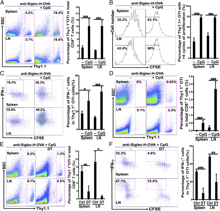
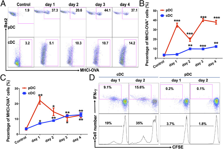
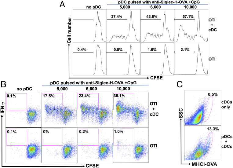
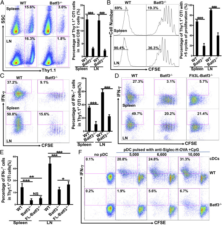
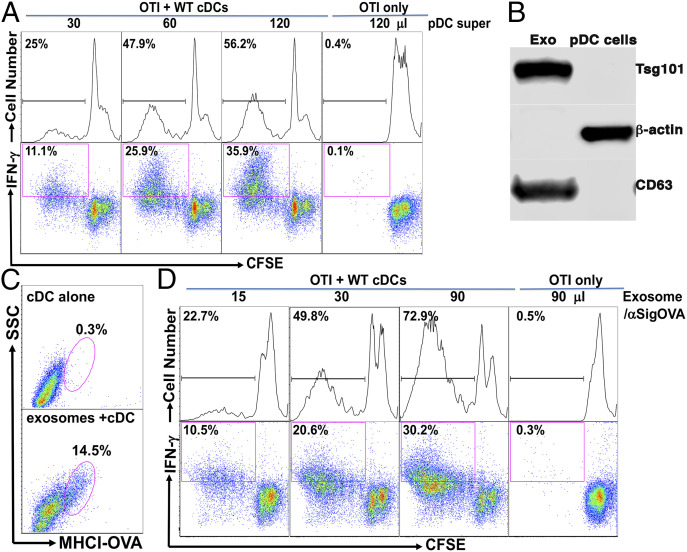
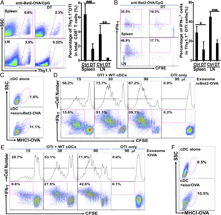
Although plasmacytoid dendritic cells (pDCs) have been shown to play a critical role in generating viral immunity and promoting tolerance to suppress antitumor immunity, whether and how pDCs cross-prime CD8 T cells in vivo remain controversial. Using a pDC-targeted vaccine model to deliver antigens specifically to pDCs, we have demonstrated that pDC-targeted vaccination led to strong cross-priming and durable CD8 T cell immunity. Surprisingly, cross-presenting pDCs required conventional DCs (cDCs) to achieve cross-priming in vivo by transferring antigens to cDCs. Taking advantage of an in vitro system where only pDCs had access to antigens, we further demonstrated that cross-presenting pDCs were unable to efficiently prime CD8 T cells by themselves, but conferred antigen-naive cDCs the capability of cross-priming CD8 T cells by transferring antigens to cDCs. Although both cDC1s and cDC2s exhibited similar efficiency in acquiring antigens from pDCs, cDC1s but not cDC2s were required for cross-priming upon pDC-targeted vaccination, suggesting that cDC1s played a critical role in pDC-mediated cross-priming independent of their function in antigen presentation. Antigen transfer from pDCs to cDCs was mediated by previously unreported pDC-derived exosomes (pDCexos), that were also produced by pDCs under various conditions. Importantly, all these pDCexos primed naive antigen-specific CD8 T cells only in the presence of bystander cDCs, similarly to cross-presenting pDCs, thus identifying pDCexo-mediated antigen transfer to cDCs as a mechanism for pDCs to achieve cross-priming. In summary, our data suggest that pDCs employ a unique mechanism of pDCexo-mediated antigen transfer to cDCs for cross-priming.

Keywords: antigen transfer; conventional dendritic cells; cross-priming; exosomes; plasmacytoid dendritic cells.


Conflict of interest statement

The authors declare no competing interest.

Figures

Fig. 1.
Fig. 1.
Targeting pDCs by anti-Siglec-H-OVA plus CpG led to strong OVA-specific CD8 T cell responses that were dependent on cDCs. (AC) WT mice were immunized with anti-Siglec-H-OVA either alone or with CpG (n = 5), and cross-priming was examined as described in Materials and Methods. (A) The percentages of Thy1.1+ OTI cells out of total CD8 T cells are depicted with representative plots on the Left and bar graph with statistics analysis on the Right. The percentages of Thy1.1+ OTI cells that had undergone more than five cycles of proliferation are depicted in B with representative histograms and bar graph with statistics analysis. The percentages of IFN-γ+ cells out of total Thy1.1+ OTI cells are shown in C with representative plots and statistics analysis in bar graph. Student’s t test was performed for AF and NS = P > 0.05%, *P < 0.05, **P < 0.01, ***P < 0.001. (D) Immunization with pDC-targeting anti-Siglec-H-OVA alone led to tolerance while immunization with anti-Siglec-H-OVA plus CpG led to strong recall responses. WT mice (n = 5) were immunized as in A and recalled at 21 d with OVA in CFA, and recall responses were examined. The percentages of Thy1.1+ OTI cells out of total CD8 T cells are depicted. (E and F) cDCs play a critical role in cross-priming upon immunization with pDC-targeting anti-Siglec-H-OVA plus CpG. CD11c-DTR→WT bone marrow chimeras (n = 3 to 5) were treated with DT or PBS on days −2, 0, and 2, and cross-priming was examined. The percentages of Thy1.1+ OTI cells out of total CD8 T cells are shown in E; and mean and SD of the percentages of IFN-γ+ cells out of total Thy1.1+ OTI cells are shown in F. Data shown are representative of at least three independent experiments.
Fig. 2.
Fig. 2.
Targeting pDCs with anti-Siglec-H-OVA plus CpG in vivo led to the expression of functional MHCI-OVA (H-2Kb-SIINFEKL) complexes on cDCs. (AC) WT mice were immunized with anti-Siglec-H-OVA plus CpG, and spleen and LNs were processed at indicated times (n = 3) and subjected to flow cytometry. pDCs and cDCs were gated as CD11cintermediate Bst2+ and CD11chigh Bst2 cells, respectively. The percentages of H-2Kb-SIINFEKL+ cells out of total pDCs and cDCs in spleens are shown in A with representative flow plots in B. The percentages of H-2Kb-SIINFEKL+ cells out of total pDCs and cDCs in LNs are depicted in C. Each time point was compared to controls. (D) cDCs but not pDCs from immunized WT mice were able to prime naive OTI CD8 T cells. pDCs and cDCs were isolated from pooled spleens of WT mice immunized as in A and then cocultured with naive OTI cells. The percentages of IFN-γ+ cells out of total Thy1.1+ OTI cells are shown (Upper), and the percentages of proliferated (CFSElow) OTI cells out of total OTI cells are shown (Lower). Data shown are representative of three or more independent experiments. *P < 0.05, **P < 0.01, and ***P < 0.001.
Fig. 3.
Fig. 3.
Antigen-loaded pDCs required the help of cDCs to prime antigen-specific CD8 T cell in vitro. (A and B) While pDCs pulsed with anti-Siglec-H-OVA plus CpG failed to prime naive OTI cells by themselves, these pDCs induced strong priming of naive OTI cells in the presence of naive cDCs. pDCs were pulsed with anti-Siglec-H-OVA plus CpG for 4 h and cocultured with naive OTI cells (1 × 10⁵ cells) either with or without bystander splenic cDCs. Percentages of proliferated CFSElow OTI are shown in A and the percentages of proliferated IFN-γ+ cells (CFSElowIFN-γ+) out of total OTI cells are shown in B. (C) pDCs pulsed with anti-Siglec-H-OVA plus CpG transferred antigens to antigen-naive cDCs. pDCs pulsed with anti-Siglec-H-OVA plus CpG were cultured with freshly isolated congenic cDCs (CD45.1 vs. CD45.2), and subjected to flow cytometry. Expression of MHCI-OVA on gated cDCs are shown. Data shown are representative of three or more independent experiments.
Fig. 4.
Fig. 4.
Batf3-depedent cDC1s played a critical role in pDC-mediated cross-priming in vivo. (AC) Batf3−/− mice exhibited significantly reduced cross-priming. WT and Batf3−/− mice (n = 5) were examined for cross-priming as in Fig. 1. The percentages of Thy1.1+ OTI cells out of total CD8 T cells were depicted with representative plots in A, Upper; the percentages of Thy1.1+ OTI cells that had undergone more than five cycles of proliferation are depicted in B; and the percentages of IFN-γ+ cells out of total Thy1.1+ OTI cells are depicted in C. (D and E) Increasing the number of DCs in Batf3−/− mice failed to restore cross-priming. WT, Batf3−/−, and Flt3L-treated Batf3−/− mice (n = 4) were examined for cross-priming as in C. The percentages of IFN-γ+ cells out of total Thy1.1+ OTI cells are shown for both spleen and LNs in D with representative plots and in E with bar graph for statistics analysis. (F) WT but not Batf3−/− cDCs were able to mediate efficient priming of OTI cells by WT pDCs in vitro. WT pDCs pulsed with anti-Siglec-H-OVA plus CpG were cocultured with naive OTI cells (1 × 10⁵ cells) and 2 × 10⁴ WT or Batf3−/− cDCs as indicated. Percentages of proliferated IFN-γ+ cells (CFSElowIFN-γ+) out of total OTI cells are shown. Data are representative of two or more independent experiments. NS = P > 0.05%, *P < 0.05, **P < 0.01, and ***P < 0.001.
Fig. 5.
Fig. 5.
Antigen-presenting pDCs conditioned cDCs to prime antigen-specific CD8 T cells by a contact-independent mechanism, likely through pDC-derived exosomes. (A) pDC-produced supernatants induced a dose-dependent priming of naive OTI cells that was dependent on the presence of cDCs. pDCs were pulsed with anti-Siglec-H-OVA plus CpG and thoroughly washed before culture. Culture supernatants were added to OTI cells in the presence or absence of naive cDCs as indicated. Percentages of total proliferated cells (CFSElow) and proliferated IFN-γ+ cells (CFSElowIFN-γ+) out of total OTI cells are shown. (B) pDCs-generated exosomes were enriched for exosome markers CD63 and Tsg101. pDCs were cultured with anti-Siglec-H-OVA and CpG, and exosomes were isolated using an Invitrogen kit. Isolated exosomes and total cell lysates were subjected to Western blot for CD63, Tsg101, and β-actin. (C) pDCs-produced exosomes transferred antigens to cDCs. cDCs were cultured either alone or with pDCs-generated exosomes and subjected to flow cytometry. Expression of MHCI-OVA (H-2Kb-SIINFEKL) on cDCs is shown. (D) pDCs-produced exosomes induced a dose-dependent priming of naive OTI cells that is dependent on the presence of cDCs. pDCs-generated exosomes were added to OTI cells in the presence or absence of naive cDCs and subjected to flow cytometry. Percentages of total proliferated cells (CFSElow) and proliferated IFN-γ+ cells (CFSElowIFN-γ+) out of total OTI cells are shown. Data shown are representative of three or more independent experiments.
Fig. 6.
Fig. 6.
pDCs treated with anti-Bst2-OVA and soluble ovalbumin protein similarly generated exosomes to cross-prime OVA-specific CD8 T cells by transferring antigens to cDCs. (A and B) cDCs play a critical role in cross-priming upon immunization with pDC-targeting anti-Bst2-OVA and CpG. CD11c-DTR→WT bone marrow chimeras (n = 4) were treated with DT or PBS on days −2, 0, and 2, and immunized with anti-Bst2-OVA plus CpG following adoptive transfer of CFSE-labeled naive OTI cells. The percentages of Thy1.1+ OTI cells out of total CD8 T cells are depicted in A, and the percentages of IFN-γ+ cells out of total Thy1.1+ OTI cells are shown in B. (C) cDCs expressed functional MHCI-OVA upon coculture with pDC-derived exosomes. cDCs were cultured alone or with exosomes from pDCs treated with anti-Bst2-OVA plus CpG, and subjected to flow cytometry. Expression of MHCI-OVA (H-2Kb-SIINFEKL) on cDCs is shown. (D) Exosomes from WT pDCs treated with anti-Bst2-OVA plus CpG induced cDC-dependent priming of naive OTI CD8 T cells. WT pDCs were cultured with anti-Bst2-OVA plus CpG and isolated exosomes were added to OTI cells in the presence or absence of naive WT cDCs. Percentages of total proliferated cells (CFSElow) and proliferated IFN-γ+ cells (CFSElowIFN-γ+) out of total OTI cells were shown. (E) Exosomes from WT pDCs treated with OVA protein plus CpG induced cDC-dependent priming of naive OTI CD8 T cells. WT pDCs were cultured with soluble OVA protein plus CpG, and isolated exosomes were cultured with naive OTI cells with or without bystander cDCs. OTI cell proliferation and differentiation are presented as in D. (F) cDCs expressed functional MHCI-OVA upon coculture with exosomes from pDCs treated with ovalbumin protein. cDCs were cultured alone or with exosomes from pDCs treated with soluble OVA protein and subjected to flow cytometry. Expression of MHCI-OVA (H-2Kb-SIINFEKL) on cDCs is shown. Data shown are representative of at least two independent experiments. *P < 0.05, **P < 0.01, and ***P < 0.001.

Similar articles

Cited by

References

    1. Steinman R. M., Some interfaces of dendritic cell biology. APMIS 111, 675–697 (2003). - PubMed
    1. Colonna M., Trinchieri G., Liu Y. J., Plasmacytoid dendritic cells in immunity. Nat. Immunol. 5, 1219–1226 (2004). - PubMed
    1. Gilliet M., Cao W., Liu Y. J., Plasmacytoid dendritic cells: Sensing nucleic acids in viral infection and autoimmune diseases. Nat. Rev. Immunol. 8, 594–606 (2008). - PubMed
    1. Swiecki M., Colonna M., The multifaceted biology of plasmacytoid dendritic cells. Nat. Rev. Immunol. 15, 471–485 (2015). - PMC - PubMed
    1. Alculumbre S. G. et al. ., Diversification of human plasmacytoid predendritic cells in response to a single stimulus. Nat. Immunol. 19, 63–75 (2018). - PubMed

Publication types

LinkOut - more resources