

Skull Base Dural Closure Using a Modified Nonpenetrating Clip Device via an Endoscopic Endonasal Approach: Technical Note
- PMID: 32879185
- PMCID: PMC7555157
- DOI: 10.2176/nmc.tn.2020-0103
Skull Base Dural Closure Using a Modified Nonpenetrating Clip Device via an Endoscopic Endonasal Approach: Technical Note
Abstract
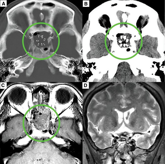
Skull base reconstruction after an endoscopic endonasal approach into the cerebrospinal fluid (CSF) space is always challenging. Various reconstructive methods are available, but no standard technique is established. This report describes the endoscopic skull base dural closure using a modified nonpenetrating clip device with shaft length of 15 cm. Six patients with an intra-suprasellar or suprasellar tumor who underwent extended endoscopic endonasal transsphenoidal surgery were targeted. For closure of the skull base dural defect after tumor removal, fascia lata was first placed as an inlay graft and was subsequently fixed with the dura using a modified nonpenetrating clip device. No CSF leakage from the closed dura with an inlay fascia lata fixed with clips was confirmed by the Valsalva maneuver. To complete skull base reconstruction, fascia lata was then positioned as an overlay graft and covered with vascularized pedicled nasoseptal flaps. Five of six patients experienced no CSF rhinorrhea postoperatively. The modified nonpenetrating clip device may achieve effective dural closure in the deep and narrow nasal cavity. We introduce this clip device technique as one of the endoscopic skull base dural closure methods.
Keywords: endoscopic skull base surgery; extended endoscopic endonasal transsphenoidal surgery; nonpenetrating titanium clip; skull base dural closure.
Conflict of interest statement
The authors declare that they have no conflict of interest.
Figures


Similar articles
-
Skull base reconstruction in the extended endoscopic transsphenoidal approach for suprasellar lesions.J Neurosurg. 2007 Oct;107(4):713-20. doi: 10.3171/JNS-07/10/0713. J Neurosurg. 2007. PMID: 17937213 Clinical Trial.
-
How I do it: shoelace watertight dural closure in extended transsphenoidal surgery.Acta Neurochir (Wien). 2015 Dec;157(12):2089-92. doi: 10.1007/s00701-015-2612-4. Epub 2015 Oct 19. Acta Neurochir (Wien). 2015. PMID: 26477503
-
Cross-reinforcing suturing and intranasal knotting for dural defect reconstruction during endoscopic endonasal skull base surgery.Acta Neurochir (Wien). 2020 Oct;162(10):2409-2412. doi: 10.1007/s00701-020-04367-w. Epub 2020 May 16. Acta Neurochir (Wien). 2020. PMID: 32418133
-
Endoscopic endonasal pituitary and skull base surgery.Neurol Med Chir (Tokyo). 2010;50(9):756-64. doi: 10.2176/nmc.50.756. Neurol Med Chir (Tokyo). 2010. PMID: 20885110 Review.
-
Cranial base repair with combined vascularized nasal septal flap and autologous tissue graft following expanded endonasal endoscopic neurosurgery.J Neurol Surg A Cent Eur Neurosurg. 2013 Mar;74(2):101-8. doi: 10.1055/s-0032-1330118. Epub 2013 Jan 14. J Neurol Surg A Cent Eur Neurosurg. 2013. PMID: 23319331 Review.
Cited by
-
Risk Factors for Postoperative Cerebrospinal Fluid Leak after Graded Multilayer Cranial Base Repair with Suturing via the Endoscopic Endonasal Approach.Neurol Med Chir (Tokyo). 2023 Feb 15;63(2):48-57. doi: 10.2176/jns-nmc.2022-0132. Epub 2022 Nov 25. Neurol Med Chir (Tokyo). 2023. PMID: 36436977 Free PMC article.
-
Creation of low cost, simple, and easy-to-use training kit for the dura mater suturing in endoscopic transnasal pituitary/skull base surgery.Sci Rep. 2023 Apr 13;13(1):6073. doi: 10.1038/s41598-023-32311-2. Sci Rep. 2023. PMID: 37055468 Free PMC article.
References
-
- Kitano M, Taneda M: Subdural patch graft technique for watertight closure of large dural defects in extended transsphenoidal surgery. Neurosurgery 54: 653–660; discussion 660-661, 2004 - PubMed
-
- Hadad G, Bassagasteguy L, Carrau RL, et al. : A novel reconstructive technique after endoscopic expanded endonasal approaches: vascular pedicle nasoseptal flap. Laryngoscope 116: 1882–1886, 2006 - PubMed
-
- Horiguchi K, Murai H, Hasegawa Y, Hanazawa T, Yamakami I, Saeki N: Endoscopic endonasal skull base reconstruction using a nasal septal flap: surgical results and comparison with previous reconstructions. Neurosurg Rev 33: 235–241, discussion 241, 2010 - PubMed
-
- Kassam AB, Thomas A, Carrau RL, et al. : Endoscopic reconstruction of the cranial base using a pedicled nasoseptal flap. Neurosurgery 63: ONS44–52; discussion ONS52–53, 2008 - PubMed
-
- Locatelli D, Rampa F, Acchiardi I, Bignami M, De Bernardi F, Castelnuovo P: Endoscopic endonasal approaches for repair of cerebrospinal fluid leaks: nine-year experience. Neurosurgery 58: ONS-246–ONS256; discussion ONS–256–ONS257, 2006 - PubMed