

A deep learning approach in diagnosing fungal keratitis based on corneal photographs
- PMID: 32879364
- PMCID: PMC7468230
- DOI: 10.1038/s41598-020-71425-9
A deep learning approach in diagnosing fungal keratitis based on corneal photographs
Abstract
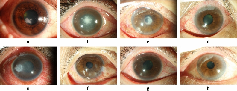
Fungal keratitis (FK) is the most devastating and vision-threatening microbial keratitis, but clinical diagnosis a great challenge. This study aimed to develop and verify a deep learning (DL)-based corneal photograph model for diagnosing FK. Corneal photos of laboratory-confirmed microbial keratitis were consecutively collected from a single referral center. A DL framework with DenseNet architecture was used to automatically recognize FK from the photo. The diagnoses of FK via corneal photograph for comparing DL-based models were made in the Expert and NCS-Oph group through a majority decision of three non-corneal specialty ophthalmologist and three corneal specialists, respectively. The average percentage of sensitivity, specificity, positive predictive value, and negative predictive value was approximately 71, 68, 60, and 78. The sensitivity was higher than that of the NCS-Oph (52%, P < .01), whereas the specificity was lower than that of the NCS-Oph (83%, P < .01). The average accuracy of around 70% was comparable with that of the NCS-Oph. Therefore, the sensitive DL-based diagnostic model is a promising tool for improving first-line medical care at rural area in early identification of FK.
Conflict of interest statement
The authors declare no competing interests.
Figures


Similar articles
-
Comparisons of deep learning algorithms for diagnosing bacterial keratitis via external eye photographs.Sci Rep. 2021 Dec 20;11(1):24227. doi: 10.1038/s41598-021-03572-6. Sci Rep. 2021. PMID: 34930952 Free PMC article.
-
Interpretable deep learning for diagnosis of fungal and acanthamoeba keratitis using in vivo confocal microscopy images.Sci Rep. 2023 Jun 2;13(1):8953. doi: 10.1038/s41598-023-35085-9. Sci Rep. 2023. PMID: 37268665 Free PMC article.
-
Differentiation of Active Corneal Infections from Healed Scars Using Deep Learning.Ophthalmology. 2022 Feb;129(2):139-146. doi: 10.1016/j.ophtha.2021.07.033. Epub 2021 Aug 2. Ophthalmology. 2022. PMID: 34352302 Free PMC article.
-
The use of in vivo confocal microscopy in fungal keratitis - Progress and challenges.Ocul Surf. 2022 Apr;24:103-118. doi: 10.1016/j.jtos.2022.03.002. Epub 2022 Mar 10. Ocul Surf. 2022. PMID: 35278721 Review.
-
An Omics Approach to Diagnosing or Investigating Fungal Keratitis.Int J Mol Sci. 2019 Jul 25;20(15):3631. doi: 10.3390/ijms20153631. Int J Mol Sci. 2019. PMID: 31349542 Free PMC article. Review.
Cited by
-
Artificial intelligence and corneal diseases.Curr Opin Ophthalmol. 2022 Sep 1;33(5):407-417. doi: 10.1097/ICU.0000000000000885. Epub 2022 Jul 12. Curr Opin Ophthalmol. 2022. PMID: 35819899 Free PMC article. Review.
-
Automatic Diagnosis of Infectious Keratitis Based on Slit Lamp Images Analysis.J Pers Med. 2023 Mar 13;13(3):519. doi: 10.3390/jpm13030519. J Pers Med. 2023. PMID: 36983701 Free PMC article.
-
A Convolutional Neural Network Using Anterior Segment Photos for Infectious Keratitis Identification.Clin Ophthalmol. 2025 Jan 7;19:73-81. doi: 10.2147/OPTH.S496552. eCollection 2025. Clin Ophthalmol. 2025. PMID: 39801570 Free PMC article.
-
Artificial Intelligence in Cornea, Refractive Surgery, and Cataract: Basic Principles, Clinical Applications, and Future Directions.Asia Pac J Ophthalmol (Phila). 2021 Jul 1;10(3):268-281. doi: 10.1097/APO.0000000000000394. Asia Pac J Ophthalmol (Phila). 2021. PMID: 34224467 Free PMC article. Review.
-
Applications of Computer Vision for Infectious Keratitis: A Systematic Review.Ophthalmol Sci. 2025 Jun 19;5(6):100861. doi: 10.1016/j.xops.2025.100861. eCollection 2025 Nov-Dec. Ophthalmol Sci. 2025. PMID: 40778364 Free PMC article. Review.
References
-
- Tew, T.B. et al. Therapeutic penetrating keratoplasty for microbial keratitis in Taiwan from 2001 to 2014. J. Formos. Med. Assoc. https://doi.org/10.1016/j.jfma.2019.09.016 (published online October 18, 2018). - PubMed
Publication types
MeSH terms
LinkOut - more resources
Full Text Sources
