Multicenter Study
. 2020 Nov;33(11):2156-2168.
doi: 10.1038/s41379-020-00661-1. Epub 2020 Sep 2.

COVID-19 pulmonary pathology: a multi-institutional autopsy cohort from Italy and New York City

Affiliations
Multicenter Study

COVID-19 pulmonary pathology: a multi-institutional autopsy cohort from Italy and New York City

Alain C Borczuk et al. Mod Pathol. 2020 Nov.

Abstract

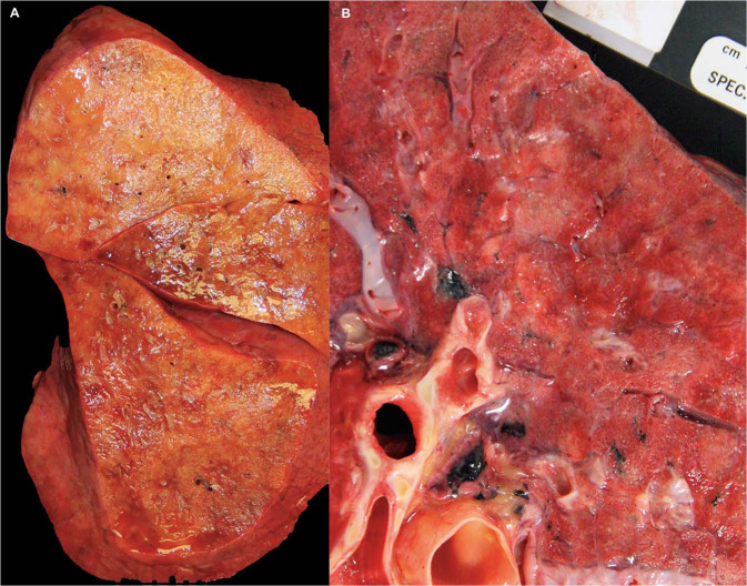
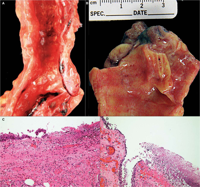
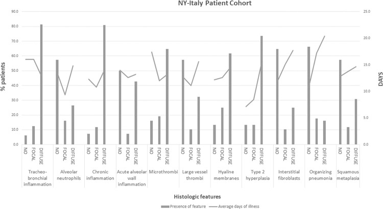
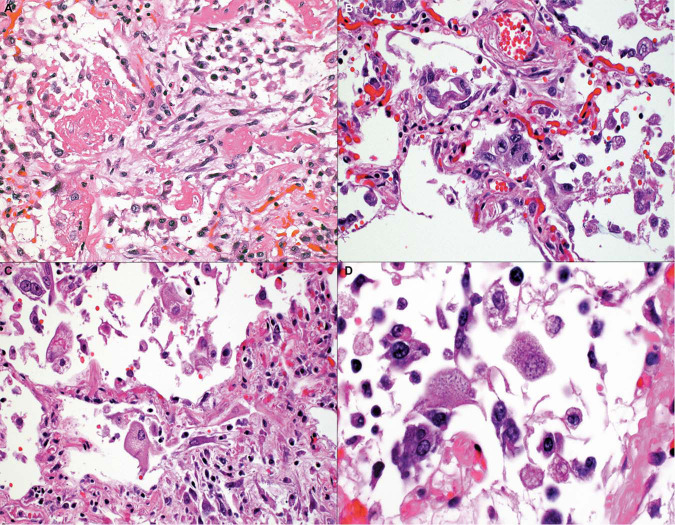
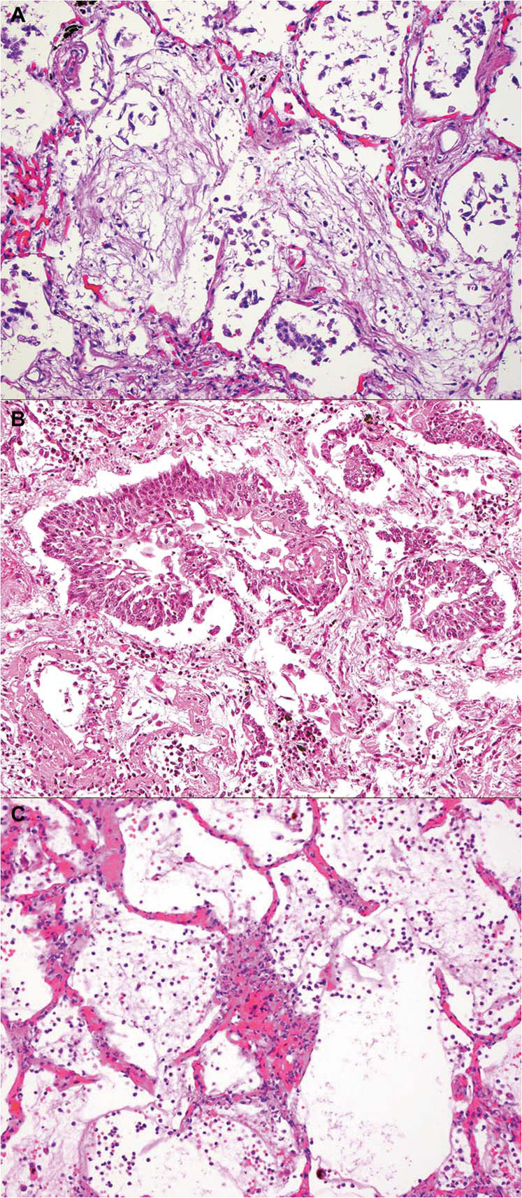
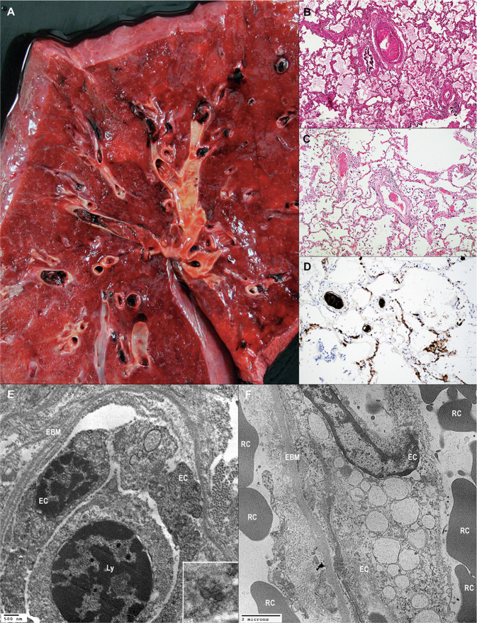
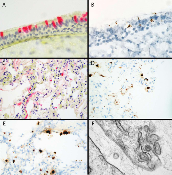
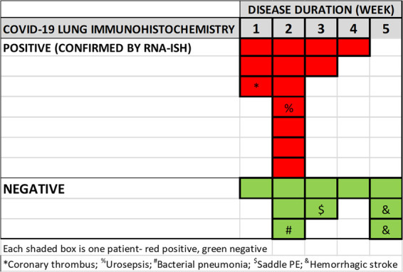
SARS-CoV-2, the etiologic agent of COVID-19, is a global pandemic with substantial mortality dominated by acute respiratory distress syndrome. We systematically evaluated lungs of 68 autopsies from 3 institutions in heavily hit areas (2 USA, 1 Italy). Detailed evaluation of several compartments (airways, alveolar walls, airspaces, and vasculature) was performed to determine the range of histologic features. The cohort consisted of 47 males and 21 females with a median age of 73 years (range 30-96). Co-morbidities were present in most patients with 60% reporting at least three conditions. Tracheobronchitis was frequently present, independent from intubation or superimposed pneumonia. Diffuse alveolar damage (DAD) was seen in 87% of cases. Later phases of DAD were less frequent and correlated with longer duration of disease. Large vessel thrombi were seen in 42% of cases but platelet (CD61 positive) and/or fibrin microthrombi were present at least focally in 84%. Ultrastructurally, small vessels showed basal membrane reduplication and significant endothelial swelling with cytoplasmic vacuolization. In a subset of cases, virus was detected using different tools (immunohistochemistry for SARS-CoV-2 viral spike protein, RNA in situ hybridization, lung viral culture, and electron microscopy). Virus was seen in airway epithelium and type 2 pneumocytes. IHC or in situ detection, as well as viable form (lung culture positive) was associated with the presence of hyaline membranes, usually within 2 weeks but up to 4 weeks after initial diagnosis. COVID-19 pneumonia is a heterogeneous disease (tracheobronchitis, DAD, and vascular injury), but with consistent features in three centers. The pulmonary vasculature, with capillary microthrombi and inflammation, as well as macrothrombi, is commonly involved. Viral infection in areas of ongoing active injury contributes to persistent and temporally heterogeneous lung damage.


Figures

Fig. 1
Fig. 1
Gross pathology, lung parenchyma. a This right lung alone weighed 1100 g and showed diffuse consolidation. b A pattern of patchy areas of gray–white consolidation was also frequently seen.
Fig. 2
Fig. 2
Tracheobronchial inflammation in COVID-19. a, b are gross photographs of trachea and main stem bronchus showing circumscribed white patches of 2.0–3.0 mm in diameter. c Microscopy of these lesions show ulceration with acute and chronic inflammation, d some with associated necrosis and fibrin. (a, b Gross photograph, Hematoxylin and eosin stain, c ×50 and d ×100).
Fig. 3
Fig. 3
Scoring of lung pathology, by days of disease. The frequency of lung findings scored as absent, focal, or diffuse by percent of total patients evaluated for that feature is shown on the left axis (bar graph) while the average days of illness for that feature (absent, focal, or diffuse) is shown on right axis (line graph).
Fig. 4
Fig. 4
Patterns of acute respiratory distress syndrome. a A combination of hyaline membranes, type 2 cell hyperplasia, and interstitial fibroblastic proliferation is shown. b Type 2 pneumocyte hyperplasia without fibroblastic proliferation. c Pneumocytes were enlarged and atypical, with (d) some pneumocytes showing multinucleated syncytial features and/or basophilic intracytoplasmic inclusions (Hematoxylin and eosin stain, a, b, c ×100, d ×150).
Fig. 5
Fig. 5
Less commonly encountered histologic features. a Young collagen with features of organizing pneumonia. b Squamous metaplasia in alveolar space. c Alveolar wall injury with neutrophils, and endothelial injury was seen in a subset of early disease duration cases (Hematoxylin and eosin stain, a, b, c ×100).
Fig. 6
Fig. 6
Vascular injury in COVID-19 lung. a A gross image showing multiple thrombi. b In addition to large thrombi, smaller caliber arteries showing fibrin thrombi. c Thrombi were seen in small arteries, including precapillary channels). d In some cases, thrombi were predominantly composed of platelets, and were also seen in the capillary bed. e The endothelial basement membrane was diffusely reduplicated. At higher magnification some putative viral particles were evident in endothelial cell cytoplasm. f Endothelial cells were swollen and showed numerous instances of cytoplasmic vacuolization. EC endothelial cell; EBM endothelial basement membrane; Ly lymphocyte; RC red cell (a—gross photograph, Hematoxylin and eosin b, c, ×100, CD61 diaminobenzidine immunohistochemistry, ×100, ef transmission electron microscopy e ×12,000, inset ×70,000, f ×6000).
Fig. 7
Fig. 7
Ancillary studies for tissue-based viral detection. a Immunohistochemistry for viral spike protein in tracheal epithelium. b RNAish for viral spike protein RNA, in same case as in (a). c Immunohistochemistry for viral spike protein in hyaline membranes and adjacent cells with (d) RNAish for viral spike protein RNA in the same area of a serial section as (c). e Lung tissue with area of new injury with background organization showing RNAish for viral spike protein in probable AT2 cells. f Electron microscopy showing a viral particle measuring 67.3 nm with electron-dense surface and the typical spikes in pneumocyte cytoplasm (a, c Alkaline phosphatase immunohistochemistry ×150, ×100, b, d, e Diaminobenzidine RNAish ×150, ×100, ×150, f Transmission electron microscopy, ×100,000).
Fig. 8
Fig. 8
COVID-19 in lung tissue by week of disease. Immunohistochemistry for viral spike protein, with positives confirmed by RNAish, tabulated by week of disease. Each box is one positive case with red positive, and green negative. The autopsy finding of associated immediate cause of death, if not solely from COVID-19 lung injury, is shown by symbols.

Similar articles

Cited by

  • A single-cell and spatial atlas of autopsy tissues reveals pathology and cellular targets of SARS-CoV-2.
    Delorey TM, Ziegler CGK, Heimberg G, Normand R, Yang Y, Segerstolpe A, Abbondanza D, Fleming SJ, Subramanian A, Montoro DT, Jagadeesh KA, Dey KK, Sen P, Slyper M, Pita-Juárez YH, Phillips D, Bloom-Ackerman Z, Barkas N, Ganna A, Gomez J, Normandin E, Naderi P, Popov YV, Raju SS, Niezen S, Tsai LT, Siddle KJ, Sud M, Tran VM, Vellarikkal SK, Amir-Zilberstein L, Atri DS, Beechem J, Brook OR, Chen J, Divakar P, Dorceus P, Engreitz JM, Essene A, Fitzgerald DM, Fropf R, Gazal S, Gould J, Grzyb J, Harvey T, Hecht J, Hether T, Jane-Valbuena J, Leney-Greene M, Ma H, McCabe C, McLoughlin DE, Miller EM, Muus C, Niemi M, Padera R, Pan L, Pant D, Pe'er C, Pfiffner-Borges J, Pinto CJ, Plaisted J, Reeves J, Ross M, Rudy M, Rueckert EH, Siciliano M, Sturm A, Todres E, Waghray A, Warren S, Zhang S, Zollinger DR, Cosimi L, Gupta RM, Hacohen N, Hide W, Price AL, Rajagopal J, Tata PR, Riedel S, Szabo G, Tickle TL, Hung D, Sabeti PC, Novak R, Rogers R, Ingber DE, Jiang ZG, Juric D, Babadi M, Farhi SL, Stone JR, Vlachos IS, Solomon IH, Ashenberg O, Porter CBM, Li B, Shalek AK, Villani AC, Rozenblatt-Rosen O, Regev A. Delorey TM, et al. bioRxiv [Preprint]. 2021 Feb 25:2021.02.25.430130. doi: 10.1101/2021.02.25.430130. bioRxiv. 2021. PMID: 33655247 Free PMC article. Preprint.
  • Clinicopathologic features of a feline SARS-CoV-2 infection model parallel acute COVID-19 in humans.
    Rudd JM, Selvan MT, Cowan S, Kao YF, Midkiff CC, Ritchey JW, Miller CA. Rudd JM, et al. bioRxiv [Preprint]. 2021 Apr 23:2021.04.14.439863. doi: 10.1101/2021.04.14.439863. bioRxiv. 2021. Update in: Viruses. 2021 Aug 05;13(8):1550. doi: 10.3390/v13081550. PMID: 33880467 Free PMC article. Updated. Preprint.
  • Plasma Metabolome Alterations Discriminate between COVID-19 and Non-COVID-19 Pneumonia.
    More TH, Mozafari B, Märtens A, Herr C, Lepper PM, Danziger G, Volk T, Hoersch S, Krawczyk M, Guenther K, Hiller K, Bals R. More TH, et al. Metabolites. 2022 Nov 2;12(11):1058. doi: 10.3390/metabo12111058. Metabolites. 2022. PMID: 36355140 Free PMC article.
  • Quantitative inspiratory-expiratory chest CT findings in COVID-19 survivors at the 6-month follow-up.
    Jia X, Han X, Cao Y, Fan Y, Yuan M, Li Y, Gu J, Zheng Y, Wang L, Qu Y, Shi H. Jia X, et al. Sci Rep. 2022 May 5;12(1):7402. doi: 10.1038/s41598-022-11237-1. Sci Rep. 2022. PMID: 35513692 Free PMC article.
  • Metabolomics Diagnosis of COVID-19 from Exhaled Breath Condensate.
    Barberis E, Amede E, Khoso S, Castello L, Sainaghi PP, Bellan M, Balbo PE, Patti G, Brustia D, Giordano M, Rolla R, Chiocchetti A, Romani G, Manfredi M, Vaschetto R. Barberis E, et al. Metabolites. 2021 Dec 6;11(12):847. doi: 10.3390/metabo11120847. Metabolites. 2021. PMID: 34940605 Free PMC article.

References

    1. Zhu N, Zhang D, Wang W, Li X, Yang B, Song J, et al. A novel coronavirus from patients with pneumonia in China, 2019. N Engl J Med. 2020;382:727–733. PMCID 7092803; PubMed PMID: 31978945. - PMC - PubMed
    1. Cucinotta D, Vanelli M. WHO declares COVID-19 a pandemic. Acta Biomed. 2020;91:157–160. PMCID 7569573; PubMed PMID: 32191675. - PMC - PubMed
    1. Huang C, Wang Y, Li X, Ren L, Zhao J, Hu Y, et al. Clinical features of patients infected with 2019 novel coronavirus in Wuhan, China. Lancet. 2020;395:497–506. PMCID 7159299; PubMed PMID: 31986264. - PMC - PubMed
    1. Zhang H, Penninger JM, Li Y, Zhong N, Slutsky AS. Angiotensin-converting enzyme 2 (ACE2) as a SARS-CoV-2 receptor: molecular mechanisms and potential therapeutic target. Intensive Care Med. 2020;46:586–590. PMCID 7079879; PubMed PMID: 32125455. - PMC - PubMed
    1. Lukassen S, Chua RL, Trefzer T, Kahn NC, Schneider MA, Muley T, et al. SARS-CoV-2 receptor ACE2 and TMPRSS2 are primarily expressed in bronchial transient secretory cells. EMBO J. 2020;39:e105114. PMCID 7232010; PubMed PMID: 32246845. - PMC - PubMed

Publication types

MeSH terms

LinkOut - more resources