. 2020 Sep-Oct;11(5):853-862.
doi: 10.1016/j.jcot.2020.07.022. Epub 2020 Jul 31.

Sacropelvic fixation techniques - Current update

Affiliations

Sacropelvic fixation techniques - Current update

Ankur Nanda et al. J Clin Orthop Trauma. 2020 Sep-Oct.

Erratum in

  • Erratum regarding previously published articles.
    [No authors listed] [No authors listed] J Clin Orthop Trauma. 2021 Aug 5;21:101558. doi: 10.1016/j.jcot.2021.101558. eCollection 2021 Oct. J Clin Orthop Trauma. 2021. PMID: 34414072 Free PMC article.

Abstract

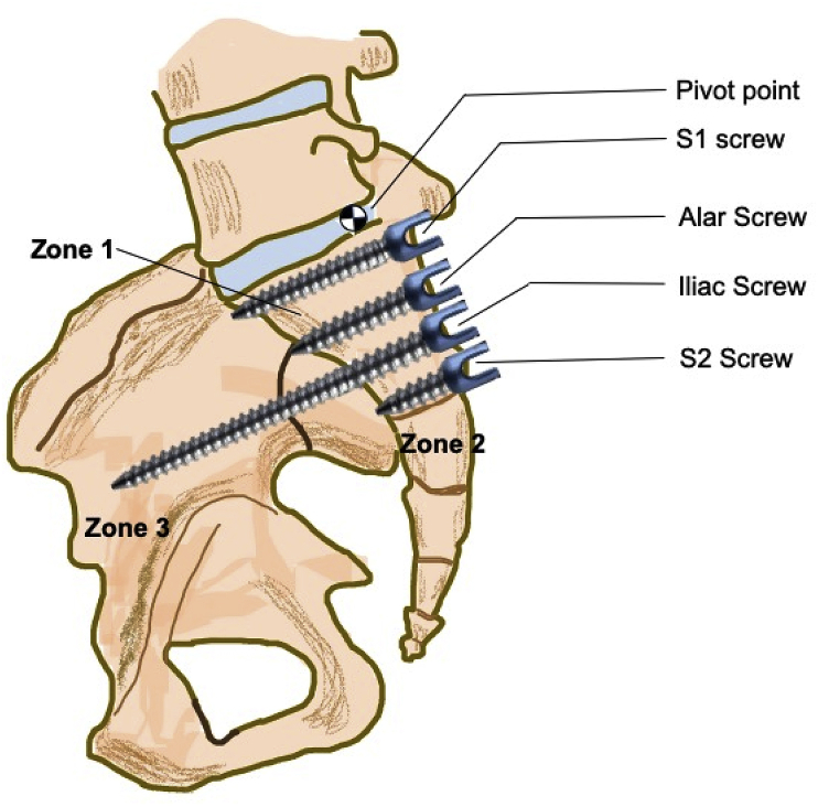
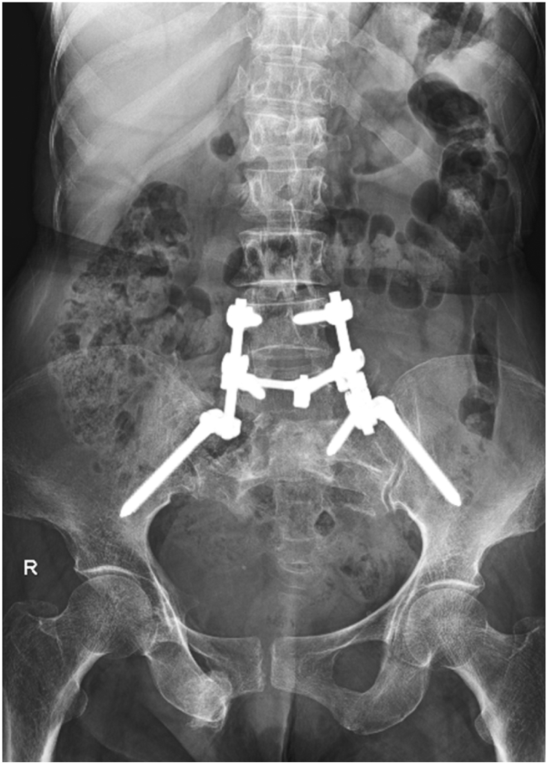
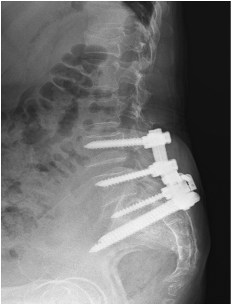
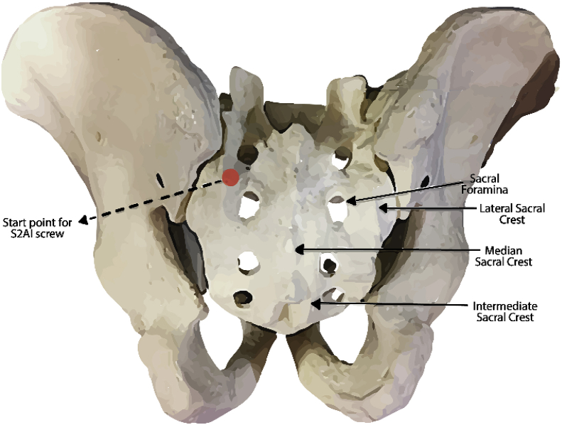
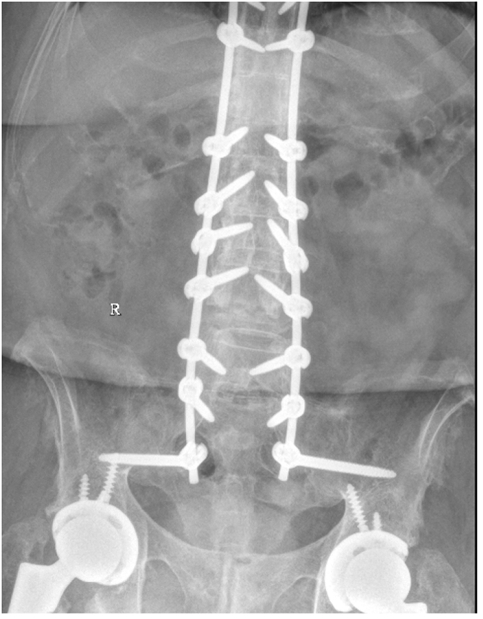
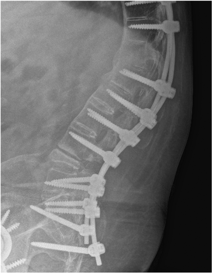
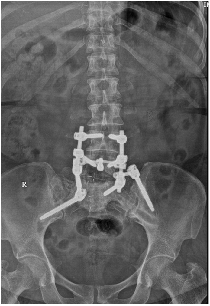
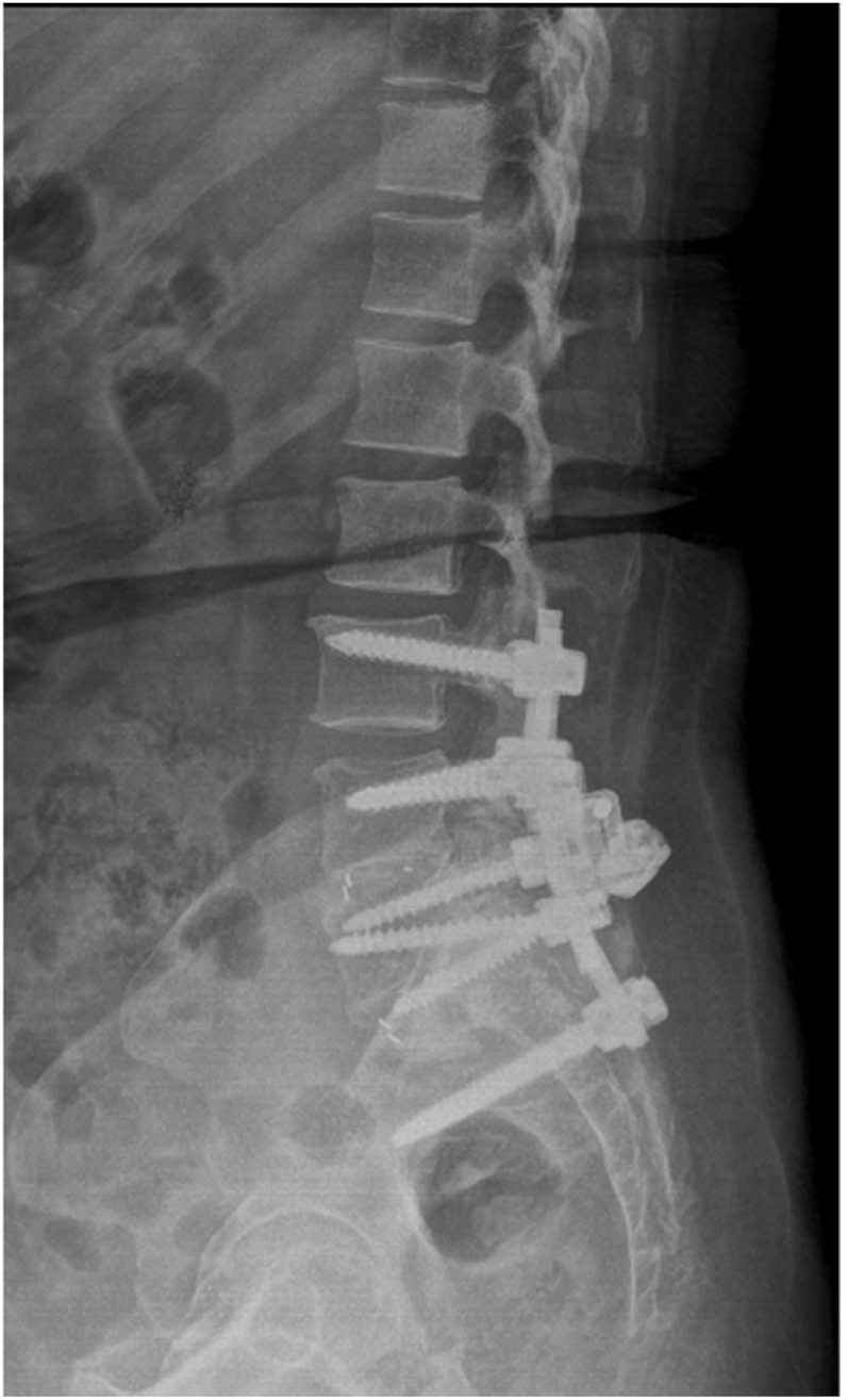
Sacropelvic is a complex junctional area owing to the complex regional anatomy and higher biomechanical stress. However extension of construct is indicated in cases with complex deformities, high grade spondylolisthesis, and complex fractures. The challenges remain which includes pseudoarthrosis and fixation failures. The fixation techniques have constantly evolved over time with better results with iliac screws and S2-alar-iliac screws. This article gives background on evolution, biomechanics, and recent update of use of robotics for sacropelvic fixation.

Keywords: Deformity; Fixation; Pelvic; Robotics; S2AI; Sacropelvic.


Figures

Fig. 1
Fig. 1
Zones of O’Brien and McCord’s pivot point.
Fig. 2
Fig. 2
Use of bilateral iliac screws for lumbosacral fusion in a 56year old female having a sacral fracture involving the right L5-S1 facet.
Fig. 3
Fig. 3
Lateral view showing bilateral iliac screws lumbosacral fusion in a 56 year old female having a sacral fracture involving the right L5-S1 facet.
Fig. 4
Fig. 4
Entry point S2AI screw (Between S1 and S2 foramen medial to the lateral sacral creast).
Fig. 5
Fig. 5
The inclination in the axial plane.
Fig. 6
Fig. 6
The trajectory is within 20 mm of sciatic notch.
Fig. 7
Fig. 7
A and B- Obturator oblique view showing screw tips within the tear drop. Source of image: Vilela MD, Braga BP, Pedrosa HAS. Fluoroscopy only for the placement of long iliac screws: A study on 14 patients. Surg Neurol Int. 2018; 9:108. Published 2018 May 25. https://doi.org/10.4103/sni.sni_59_18.
Fig. 8
Fig. 8
Steps of insertion of S2AI screws. A- Gearshift technique in which to avoid the anterior cortex breach, the probe is rotated. B- Once no breach is confirmed, k wire is inserted. C- Tapping and size of screw is determined. D- Finally, the screw is inserted.
Fig. 9
Fig. 9
Final position of S2AI screws.
Fig. 10
Fig. 10
Intra op navigation guidance and trajectory for S2AI screws Source- Phan, Kevin & Li, Julian & Giang, Gloria & Teng, Ian & Phan, Steven & Chang, Nicholas & Mobbs, Ralph. (2017). Technical note A novel technique for placement of sacro-alar-iliac (S2AI) screws by K-wire insertion using intraoperative navigation. Journal of Clinical Neuroscience. 45. 10.1016/j.jocn.2017.08.049.
Fig. 11
Fig. 11
3D reconstruction of a Traumatic sacral fracture in a 39year old female with left sacroiliac dissociation.
Fig. 12
Fig. 12
The patient was treated with a Lumbosacral construct with S2AI screws.
Fig. 13
Fig. 13
A 38/M with Ankylosing spondylitis treated with PSO at L4 for kyphotic deformity correction with horizontal gaze. The construct is supplemented with S2AI screws for better stability.
Fig. 14
Fig. 14
Lateral view showing S2AI screws in the same case.
Fig. 15
Fig. 15
An example of Sacropelvic fusion in a case of L5-S1 pseudarthrosis using left iliac screw and right sided S2AI screw for better stability of the construct.
Fig. 16
Fig. 16
Lateral view with left iliac screw and right sided S2AI screw.
Fig. 17
Fig. 17
Dual outer diameter screws.
Fig. 18
Fig. 18
Closed head DOD polyaxial, Monoaxial, Angled Monoaxial screws.

References

    1. Goodwin CR, Kosztowski TA, Elder BD, et al. Complex lumbosacropelvic fixation techniques. In: Steinmetz MP, Benzel EC, editors. Benzel’s Spine Surgery: Techniques, Complication Avoidance, and Management. Philadelphia, PA: Elsevier; 2017760e767.e2.
    1. Panjabi M.M., White A.A., III Basic biomechanics of the spine. Neurosurgery. 1980;7:76e93. - PubMed
    1. Dietrich M., Kurowski P. The importance of mechanical factors in the etiology of spondylolysis. A model analysis of loads and stresses in human lumbar spine. Spine (Phila Pa 1976) 1985;10:532e42. - PubMed
    1. Colombini D., Occhipinti E., Grieco A. Estimation of lumbar disc areas by means of anthropometric parameters. Spine (Phila Pa 1976) 1989;14:51e5. - PubMed
    1. Zheng Y., Lu W.W., Zhu Q. Variation in bone mineral density of the sacrum in young adults and its significance for sacral fixation. Spine (Phila Pa 1976) 2000;25:353e7. - PubMed