

Prevalence of cervical spine instability among Rheumatoid Arthritis patients in South Iraq
- PMID: 32879574
- PMCID: PMC7452176
- DOI: 10.1016/j.jcot.2019.06.013
Prevalence of cervical spine instability among Rheumatoid Arthritis patients in South Iraq
Abstract
Aim of the work: This study analysed the prevalence of cervical spine instability in Rheumatoid Arthritis (RA) patients following at a single centre in Basrah.
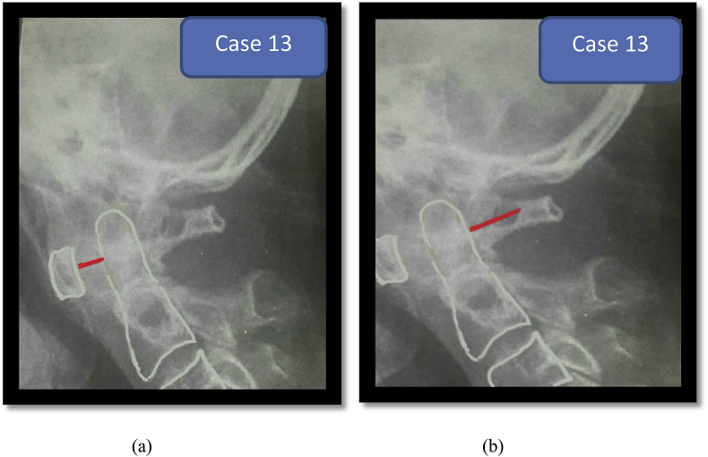
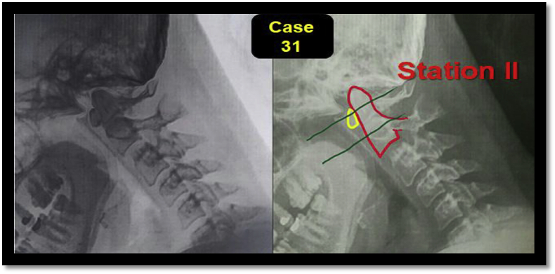
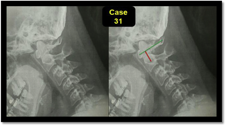
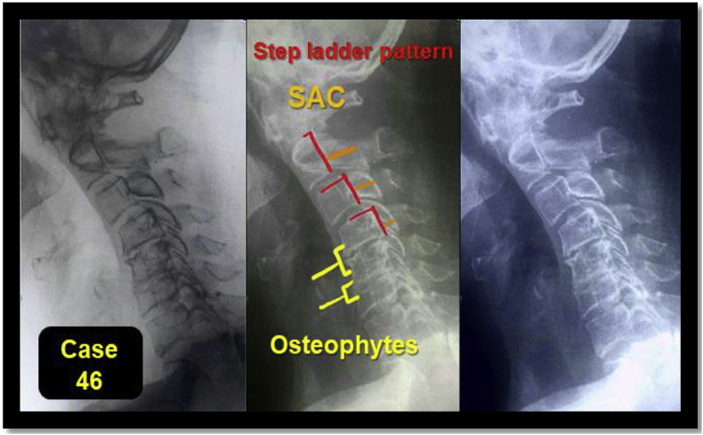
Patients and methods: Data were collected directly from patients through cervical spine examinations. Each patient was sent for dynamic (flexion and extension) lateral cervical radiographic imaging to assess the presence of atlantoaxial subluxation (AAS), superior migration of the odontoid (SMO) and sub-axial subluxation (SAS). Patients with positive radiographic findings were sent for MRI scans of the cervical spine to assess neurological compression.
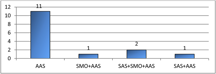
Results: The prevalence rate of cervical spine instability in RA was 15/203 (7.4%) of the total sample, occurring primarily in patients of 37-65 years old (mean: 48 ± 8.9 years), were 3/15 (20%) aymptomatic. The majority (60%) being at the moderate stage of the disease activity (using a Clinical Disease Activity Index [CDAI). In terms of type of cervical spine involvement, isolated AAS was found to have the highest occurrence (73.3%), followed by combined SAS and SMO (13.3%), combined AAS and SMO (6.7%), and combined AAS and SAS (6.7%). A significant relationship was found between the type of cervical spine involvement in RA and a disease onset duration, disease activity, body mass index and peripheral erosion with P value < 0.05.
Conclusion: Cervical spine subluxation in RA patients may be asymptomatic It is therefore essential to obtain a dynamic radiographic image of the cervical spine in order to diagnose cervical spine involvement and protect the patient from severe outcomes.The clinical trial registration number included in a the official document from Ministry of Higher Education and Science Research/Basrah University/Faculty of Medicine to Basrah Health Directorate/Research and Development Division is 72/3588 in 7 Jan 2017.
Keywords: Cervical spine instability; Iraq; Rheumatoid arthritis.
© 2019 Delhi Orthopedic Association. All rights reserved.
Figures
Similar articles
-
Recurrence of cervical spine instability in rheumatoid arthritis following previous fusion: can disease progression be prevented by early surgery?J Rheumatol. 1992 Sep;19(9):1364-70. J Rheumatol. 1992. PMID: 1433002
-
Accelerated development of cervical spine instabilities in rheumatoid arthritis: a prospective minimum 5-year cohort study.PLoS One. 2014 Feb 18;9(2):e88970. doi: 10.1371/journal.pone.0088970. eCollection 2014. PLoS One. 2014. PMID: 24558457 Free PMC article.
-
Radiological evaluation of cervical spine involvement in rheumatoid arthritis.Neurosurg Focus. 2015 Apr;38(4):E4. doi: 10.3171/2015.1.FOCUS14664. Neurosurg Focus. 2015. PMID: 25828498 Review.
-
[Population distribution and clinical characteristics in rheumatoid arthritis patients with cervical spine instability].Beijing Da Xue Xue Bao Yi Xue Ban. 2020 Dec 18;52(6):1034-1039. doi: 10.19723/j.issn.1671-167X.2020.06.008. Beijing Da Xue Xue Bao Yi Xue Ban. 2020. PMID: 33331310 Free PMC article. Chinese.
-
Cervical spine manifestations of rheumatoid arthritis: a review.Neurosurg Rev. 2021 Aug;44(4):1957-1965. doi: 10.1007/s10143-020-01412-1. Epub 2020 Oct 10. Neurosurg Rev. 2021. PMID: 33037539 Review.
Cited by
-
Risk factors for cervical instability in rheumatoid arthritis: a meta-analysis.Arch Med Sci. 2024 Mar 5;20(2):375-383. doi: 10.5114/aoms/173494. eCollection 2024. Arch Med Sci. 2024. PMID: 38757018 Free PMC article. Review.
References
-
- Prentice A.M., Jebb S.A. Beyond body mass index. Obes Rev. 2001;2(3):141–147. - PubMed
-
- Yurube T., Sumi M., Nishida K. Progression of cervical spine instabilities in rheumatoid arthritis: a prospective cohort study of outpatients over 5 years. Spine. 2011;36(8):647–653. - PubMed
-
- Feipel V., Dugailly P.M., Rooze M. Cinématique du rachis vertébral. In: Argenson C.L., Dosch J.C., Lemaire V., Bard H., Laredo J.D., editors. Imagerie du rachis cervical (GETROA opus XXVII) Sauramps médical; Montpellier: 2000. pp. 35–42.
-
- Bland J., Boushey D. Anatomy and physiology of the cervical spine. Semin Arthritis Rheum. 1990;20:1–20. - PubMed
-
- Dreyfus P., Ghezail M., Boissier M.C. Pathologie de l’apophyse odontoïde. In: Sèze S de, Ryckewaert A., Kahn M.F., Kuntz D., Dryll D., Guérin C., editors. L’actualité Rhumatologique 1992. Expansion scientifique française; Paris: 1992. pp. 88–99.
LinkOut - more resources
Full Text Sources
Research Materials
Miscellaneous
