. 2020 Jun 7;10(36):21464-21472.
doi: 10.1039/d0ra01920h. Epub 2020 Jun 4.

Photo-initiated rupture of azobenzene micelles to enable the spectroscopic analysis of antimicrobial peptide dynamics

Affiliations

Photo-initiated rupture of azobenzene micelles to enable the spectroscopic analysis of antimicrobial peptide dynamics

Matthew G Roberson et al. RSC Adv. .

Abstract

Antimicrobial peptides (AMPs) show promise for the treatment of bacterial infections, but many have undesired hemolytic activities. The AMP MP1 not only has broad spectrum bactericidal activity, but has been shown to have antitumor activity. The interaction between AMPs and cellular membranes gives rise to a peptide's cell-specificity and activity. However, direct analysis of the biophysical interactions between peptides and membrane is complex, in part due to the nature of membrane environments as well as structural changes in the peptide that occurs upon binding to the membrane. In order to investigate the interplay between cell selectivity, activity, and secondary structural changes involved in antimicrobial peptide activity, we sought to implement photolizable membrane mimics to assess the types of information available from infrared spectroscopic measurements that follow from photoinitiated peptide dynamics. Azo-surfactants (APEG) form micelles containing a photolizable azobenzene core, which upon irradiation can induce membrane deformation resulting in breakdown of micelles. Spectroscopic analysis of membrane deformation may provide insights into the physical behavior associated with unfolding and dissociation of antimicrobial peptides within a membrane environment. Herein, we synthesized and characterized two new azo-surfactants, APEGTMG and APEGNEt2MeI. Furthermore, we demonstrate the viability of azosurfactants as membrane mimics by examining both the membrane binding and dissociation induced secondary structural changes of the antimicrobial peptide, MP1.


Conflict of interest statement

There are no conflicts to declare.

Figures

Scheme 1
Scheme 1. Synthesis of APEG derivatives APEGTMG and APEGNEt2MeI. (a) TsCl, NEt3, DCM, 0 °C – rt, 18 h, 83% yield; (b) TMG, K2CO3, THF, reflux, 48 h, 91% yield; (c) HNEt2, K2CO3, THF, reflex, 48 h, 91% yield; (d) MeI, THF, reflux, 24 h, 85% yield.
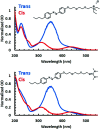
Fig. 1
Fig. 1. UV-Vis spectra of APEGNEt2MeI (top) and APEGTMG (bottom) demonstrating the electronic transitions associated with each isomer, trans (blue) and cis (red), obtained prior to irradiation with 365 nm and upon irradiating samples with 365 nm, respectively.
Fig. 2
Fig. 2. CMCs of the trans and cis isomers of the APEG surfactants were calculated by monitoring the changes in chemical shifts (depicted here as the chemical shifts of the proton labelled as “J”) as a function of surfactant concentration using 1H NMR.
Fig. 3
Fig. 3. Linear IR spectra of MP1/D2O (blue solid), MP1 with trans APEGTMG micelles (red dashed). The green difference spectrum is associated with changes in MP1 structure upon incorporation of trans APEGTMG micelles.
Fig. 4
Fig. 4. Linear IR spectra (insets) of MP1 (4.8 mM) in APEGTMG (1.74 mM) with trans APEGTMG micelles after initial preparation (t = 0 h) (black dashed inset), trans micelles after 24 h with no irradiation (red solid inset), and cis micelles after photoconversion (blue dashed inset) were collected. The pink difference spectrum (top) shows an increase of the bound peptide population (1651 cm−1) after 24 h equilibration period. The orange difference spectrum (bottom) shows the loss of the bound peptide population and return to the original state upon photoconversion (365 nm irradiation), which causes the micelles to rupture.
Fig. 5
Fig. 5. Fluorescence spectra (top) and normalized fluorescence spectra (bottom) of TR-CMP1 (47 nM) were collected in the presence (red) and absence (blue) of TMG micelles (10 mM), to assess the ability of peptide binding towards APEGTMG micelles. Normalized fluorescence spectra were normalized to the maximum emission of each individual spectrum.

References

    1. Brown K. L. Hancock R. E. W. Curr. Opin. Immunol. 2006;18:24–30. doi: 10.1016/j.coi.2005.11.004. - DOI - PubMed
    1. Hancock R. E. W. Diamond G. Trends Microbiol. 2000;8:402–410. doi: 10.1016/S0966-842X(00)01823-0. - DOI - PubMed
    1. Hoskin D. W. Ramamoorthy A. Biochim. Biophys. Acta. 2008;1778:357–375. doi: 10.1016/j.bbamem.2007.11.008. - DOI - PMC - PubMed
    1. Huang H. W. Biochemistry. 2000;39:8347–8352. - PubMed
    1. Tucker M. J. Oyola R. Gai F. J. Phys. Chem. B. 2005;109:4788–4795. doi: 10.1021/jp044347q. - DOI - PubMed