Diagnosis of Collagen Type III Glomerulopathy Using Picrosirius Red and PASH/Masson's Trichrome Stain
- PMID: 32880237
- PMCID: PMC8942299
- DOI: 10.1177/0300985820934111
Diagnosis of Collagen Type III Glomerulopathy Using Picrosirius Red and PASH/Masson's Trichrome Stain
Abstract
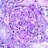
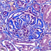
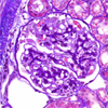
Canine collagen type III glomerulopathy (Col3GP) is a rare juvenile nephropathy in which irregular type III collagen fibrils and fibronectin accumulate in glomerular capillary walls and the mesangium. Necropsy findings were reviewed from 5 puppies diagnosed with Col3GP at 6 to 18 weeks of age. Histologically, with hematoxylin and eosin stain, the glomerular capillary walls and mesangium were diffusely and globally expanded by homogeneous pale eosinophilic material. Ultrastructurally, the subendothelial zone and mesangium were expanded by fibronectin and cross-banded collagen type III fibrils, diagnostic of Col3GP. Two additional stains were employed to identify the material within glomeruli as fibrillar collagen using light microscopy. In all 5 cases, the material was red with picrosirius red and birefringent under polarized light, and was blue with periodic acid-Schiff/hematoxylin/trichrome (PASH/TRI), thereby identifying it as fibrillar collagen. Based on these unique staining characteristics with picrosirius red and PASH/TRI, Col3GP may be reliably diagnosed with light microscopy alone.
Keywords: collagen type III; collagenofibrotic glomerulopathy; dogs; kidney; picrosirius red; renal; trichrome.
Conflict of interest statement
Declaration of Conflicting Interests
The author(s) declared no potential conflicts of interest with respect to the research, authorship, and/or publication of this article.
Figures


Similar articles
-
Additional Value of Second Harmonic Generation Microscopy in the Diagnosis of Feline Collagen Type III Glomerulopathy.J Comp Pathol. 2021 Oct;188:37-43. doi: 10.1016/j.jcpa.2021.08.005. Epub 2021 Sep 24. J Comp Pathol. 2021. PMID: 34686276
-
Collagenofibrotic glomerulonephropathy with fibronectin deposition in a dog.Vet Pathol. 2009 Jul;46(4):688-92. doi: 10.1354/vp.08-VP-0272-S-CR. Epub 2009 Mar 9. Vet Pathol. 2009. PMID: 19276051
-
Glomerular Collagen V Codeposition and Hepatic Perisinusoidal Collagen III Accumulation in Canine Collagen Type III Glomerulopathy.Vet Pathol. 2015 Nov;52(6):1134-41. doi: 10.1177/0300985814560237. Epub 2014 Dec 8. Vet Pathol. 2015. PMID: 25487411
-
Collagenofibrotic glomerulopathy: clinicopathologic overview of a rare glomerular disease.Am J Kidney Dis. 2007 Apr;49(4):499-506. doi: 10.1053/j.ajkd.2007.01.020. Am J Kidney Dis. 2007. PMID: 17386317 Review.
-
Collagenofibrotic glomerulopathy: A case of glomerular deposition disease in the Indian subcontinent and review of the literature.Saudi J Kidney Dis Transpl. 2020 May-Jun;31(3):681-686. doi: 10.4103/1319-2442.289454. Saudi J Kidney Dis Transpl. 2020. PMID: 32655054 Review.
Cited by
-
Malakoplakia in the Urinary Bladder of 4 Puppies.Vet Pathol. 2021 Jul;58(4):699-704. doi: 10.1177/03009858211009779. Epub 2021 Apr 23. Vet Pathol. 2021. PMID: 33888013 Free PMC article.
-
Mannan-Binding Lectin Is Associated with Inflammation and Kidney Damage in a Mouse Model of Type 2 Diabetes.Int J Mol Sci. 2024 Jun 29;25(13):7204. doi: 10.3390/ijms25137204. Int J Mol Sci. 2024. PMID: 39000309 Free PMC article.
References
-
- Adachi K, Mori T, Ito T, et al. Collagenofibrotic glomerulonephropathy in a cynomolgus macaque (Macaca fascicularis). Vet Pathol. 2005;42(5):669–674. - PubMed
-
- Alchi B, Nishi S, Narita I, et al. Collagenfibrotic glomerulopathy: clinicopathologic overview of a rare glomerular disease. Am J Kidney Dis. 2007;49(4):499–506. - PubMed
-
- Brodsky SV, Albawardi A, Satoskar AA, et al. When one plus one equals more than two—a novel stain for renal biopsies is a combination of two classical stains. Histol Histopathol. 2010;25(11):1379–1383. - PubMed
-
- Cianciolo RE, Brown C, Mohr C, et al. Atlas of Renal Lesions in Proteinuric Dogs. Press Books; 2019.
MeSH terms
Substances
Grants and funding
LinkOut - more resources
Full Text Sources
Medical

