Review
. 2020 Sep 3;20(11):49.
doi: 10.1007/s11910-020-01072-0.

Congenital Zika Virus Infection: a Review with Emphasis on the Spectrum of Brain Abnormalities

Affiliations
Review

Congenital Zika Virus Infection: a Review with Emphasis on the Spectrum of Brain Abnormalities

Leão Vhp et al. Curr Neurol Neurosci Rep. .

Abstract

Purpose of review: In 2016, the World Health Organization declared the Zika virus (ZIKV) outbreak a Public Health Emergency of International Concern following a cluster of associated neurological disorders and neonatal malformations. Our aim is to review the clinical and neuroimaging findings seen in congenital Zika syndrome.

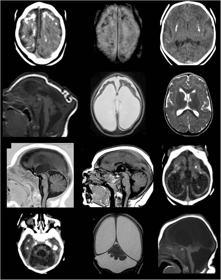
Recent findings: ZIKV injures neural progenitor cells in the hippocampus, a brain region important for learning, memory, cognition, and emotion/stress response. Positron emission tomography has revealed global neuroinflammation in ZIKV infection in animal models. Congenital Zika syndrome is associated with a spectrum of brain abnormalities, including microcephaly, parenchymal calcifications, malformations of cortical development and defective neuronal migration, corpus callosum abnormalities, ventriculomegaly, and brainstem and cerebellar abnormalities.

Keywords: Microcephaly; Neuroimaging; Zika virus; Zika virus infection.


Conflict of interest statement

Leão VHP, Aragão MM, Pinho RS, Hazin AN, Paciorkowski AR, Penalva de Oliveira AC, and Masruha, MR each declare no potential conflicts of interest.

Figures

Fig. 1
Fig. 1
Brain images of infants diagnosed with CZS. CT scan slices: images a, c, i, j, and k. MRI images: b, d, e, f, g, h, k, and l. a (CT) and b (MRI): coarse and punctate calcifications at the cortico-subcortical junction, predominating in the frontal lobes. c: basal ganglia calcifications. d: marked microcephaly with redundant scalp skin in the occipital region. e: marked dilatation of supratentorial ventricular system, simplified gyral pattern of the brain, and prominence of the subarachnoid space in the frontal regions. f: malformations of cortical development compatible with polymicrogyria in the frontal lobes with sparing of posterior cortical areas. g: thin/hypoplastic corpus callosum. h: short corpus callosum with dysgenetic aspect. i and j: tetraventricular dilatation, simplified gyral pattern of the brain, cerebellar and pons hypoplasia, and cortico-subcortical and periventricular calcifications. k: asymmetric cerebellar hypoplasia with greater involvement of the right hemisphere, marked dilatation of the lateral ventricles with thinning of cerebral mantle, and prominence of cisterna magna. l: severe cerebellar hypoplasia

References

    1. Campos GS, Bandeira AC, Sardi SI. Zika virus outbreak, Bahia, Brazil. Emerg Infect Dis. 2015;21(10):1885–1886. - PMC - PubMed
    1. Newman C, Friedrich TC, O’Connor DH. Macaque monkeys in Zika virus research: 1947-present. Curr Opin Virol. 2017;25:34–40. - PMC - PubMed
    1. Christofferson RC. Zika virus emergence and expansion: lessons learned from dengue and chikungunya may not provide all the answers. Am J Trop Med Hyg. 2016;95(1):15–18. - PMC - PubMed
    1. Musso D, Gubler DJ. Zika virus. Clin Microbiol Rev. 2016;29(3):487–524. - PMC - PubMed
    1. Hussain A, Ali F, Latiwesh OB, Hussain S. A comprehensive review of the manifestations and pathogenesis of Zika virus in neonates and adults. Cureus. 2018;10(9):e3290. - PMC - PubMed

MeSH terms