

QiShenYiQi pill improves the reparative myocardial fibrosis by regulating autophagy
- PMID: 32881330
- PMCID: PMC7576289
- DOI: 10.1111/jcmm.15695
QiShenYiQi pill improves the reparative myocardial fibrosis by regulating autophagy
Abstract
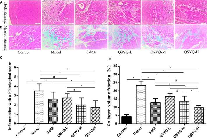
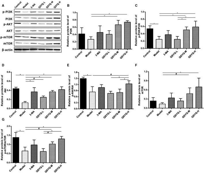
QiShenYiQi pill (QSYQ), a traditional Chinese medicine, is well known for improving the myocardial remodelling, but the dose-effect relationship of its intervention in the reparative myocardial fibrosis is still unclear. We investigated the effect of QSYQ on the reparative myocardial fibrosis in cardiac myosin-induced rats and explored its mechanism of action by regulating autophagy. The results indicated that QSYQ increased LVEF and LVFS, and decreased the LVEDD, LVESD, HMI, LVMI, myocardial inflammation histology score, and collagen volume fraction in a dose-dependent manner. In addition, QSYQ declined the number of autophagosomes, down-regulated the expression of myocardial Beclin-1 and LC3B, up-regulated the expression of myocardial p62 and increased the ratios of myocardial p-PI3K/PI3K, p-Akt/Akt and p-mTOR/mTOR. We provided evidence for that QSYQ could inhibit excessive myocardial autophagy by regulating the PI3K/Akt-mTOR pathway and can be a potential therapeutic approach in treating the cardiovascular diseases such as myocarditis and dilated cardiomyopathy.
Keywords: PI3K/Akt-mTOR pathway; autophagy; reparative myocardial fibrosis; traditional Chinese medicine.
© 2020 The Authors. Journal of Cellular and Molecular Medicine published by Foundation for Cellular and Molecular Medicine and John Wiley & Sons Ltd.
Conflict of interest statement
The authors declare no competing interests.
Figures


Similar articles
-
QiShenYiQi pill activates autophagy to attenuate reactive myocardial fibrosis via the PI3K/AKT/mTOR pathway.Aging (Albany NY). 2021 Feb 11;13(4):5525-5538. doi: 10.18632/aging.202482. Epub 2021 Feb 11. Aging (Albany NY). 2021. PMID: 33582656 Free PMC article.
-
Choline Inhibits Ischemia-Reperfusion-Induced Cardiomyocyte Autophagy in Rat Myocardium by Activating Akt/mTOR Signaling.Cell Physiol Biochem. 2018;45(5):2136-2144. doi: 10.1159/000488049. Epub 2018 Mar 7. Cell Physiol Biochem. 2018. PMID: 29533930
-
Lycium barbarum polysaccharide attenuates diabetic testicular dysfunction via inhibition of the PI3K/Akt pathway-mediated abnormal autophagy in male mice.Cell Tissue Res. 2018 Dec;374(3):653-666. doi: 10.1007/s00441-018-2891-1. Epub 2018 Aug 2. Cell Tissue Res. 2018. PMID: 30073544
-
Hydrogen sulfide ameliorates doxorubicin‑induced myocardial fibrosis in rats via the PI3K/AKT/mTOR pathway.Mol Med Rep. 2021 Apr;23(4):299. doi: 10.3892/mmr.2021.11938. Epub 2021 Mar 2. Mol Med Rep. 2021. PMID: 33649809 Free PMC article.
-
Chinese Medicine for the Treatment of Liver Cirrhosis: The Mechanism of Cellular Autophagy.Am J Chin Med. 2025;53(2):409-433. doi: 10.1142/S0192415X25500168. Epub 2025 Mar 12. Am J Chin Med. 2025. PMID: 40070295 Review.
Cited by
-
Integrative applications of network pharmacology and molecular docking: An herbal formula ameliorates H9c2 cells injury through pyroptosis.J Ginseng Res. 2023 Mar;47(2):228-236. doi: 10.1016/j.jgr.2022.03.003. Epub 2022 Mar 28. J Ginseng Res. 2023. PMID: 36926601 Free PMC article.
-
Huangqi Shengmai Yin Ameliorates Myocardial Fibrosis by Activating Sirtuin3 and Inhibiting TGF-β/Smad Pathway.Front Pharmacol. 2021 Aug 13;12:722530. doi: 10.3389/fphar.2021.722530. eCollection 2021. Front Pharmacol. 2021. PMID: 34483934 Free PMC article.
-
Hydrogen sulfide alleviates hypothyroidism-induced myocardial fibrosis in rats through stimulating autophagy and inhibiting TGF-β1/Smad2 pathway.Korean J Physiol Pharmacol. 2023 Jan 1;27(1):1-8. doi: 10.4196/kjpp.2023.27.1.1. Korean J Physiol Pharmacol. 2023. PMID: 36575928 Free PMC article.
-
Traditional Chinese Medicine Treating Dilated Cardiomyopathy: A Literature Review.Int J Gen Med. 2025 Aug 8;18:4311-4325. doi: 10.2147/IJGM.S524477. eCollection 2025. Int J Gen Med. 2025. PMID: 40809953 Free PMC article. Review.
-
Review of Chinese medicine intervention in PI3K/AKT pathway to regulate fibrosis.Medicine (Baltimore). 2025 Jul 11;104(28):e42957. doi: 10.1097/MD.0000000000042957. Medicine (Baltimore). 2025. PMID: 40660593 Free PMC article. Review.
References
-
- Knaapen P, Götte MJW, Paulus WJ, et al. Does myocardial fibrosis hinder contractile function and perfusion in idiopathic dilated cardiomyopathy? PET and MR imaging study. Radiology. 2006;240(2):380‐388. - PubMed
-
- Rubiś P, Totoń‐Żurańska J, Wiśniowska‐Śmiałek S, et al. Relations between circulating microRNAs (miR‐21, miR‐26, miR‐29, miR‐30 and miR‐133a), extracellular matrix fibrosis and serum markers of fibrosis in dilated cardiomyopathy. Int J Cardiol. 2017;231:201‐206. - PubMed
-
- Komajda M, Jais JP, Reeves F, et al. Factors predicting mortality in idiopathic dilated cardiomyopathy. Eur Heart J. 1990;11(9):824‐831. - PubMed
Publication types
MeSH terms
Substances
LinkOut - more resources
Full Text Sources
Miscellaneous
