Treatment with galectin-1 improves myogenic potential and membrane repair in dysferlin-deficient models
- PMID: 32881965
- PMCID: PMC7470338
- DOI: 10.1371/journal.pone.0238441
Treatment with galectin-1 improves myogenic potential and membrane repair in dysferlin-deficient models
Abstract
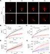
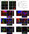
Limb-girdle muscular dystrophy type 2B (LGMD2B) is caused by mutations in the dysferlin gene, resulting in non-functional dysferlin, a key protein found in muscle membrane. Treatment options available for patients are chiefly palliative in nature and focus on maintaining ambulation. Our hypothesis is that galectin-1 (Gal-1), a soluble carbohydrate binding protein, increases membrane repair capacity and myogenic potential of dysferlin-deficient muscle cells and muscle fibers. To test this hypothesis, we used recombinant human galectin-1 (rHsGal-1) to treat dysferlin-deficient models. We show that rHsGal-1 treatments of 48 h-72 h promotes myogenic maturation as indicated through improvements in size, myotube alignment, myoblast migration, and membrane repair capacity in dysferlin-deficient myotubes and myofibers. Furthermore, increased membrane repair capacity of dysferlin-deficient myotubes, independent of increased myogenic maturation is apparent and co-localizes on the membrane of myotubes after a brief 10min treatment with labeled rHsGal-1. We show the carbohydrate recognition domain of Gal-1 is necessary for observed membrane repair. Improvements in membrane repair after only a 10 min rHsGal-1treatment suggest mechanical stabilization of the membrane due to interaction with glycosylated membrane bound, ECM or yet to be identified ligands through the CDR domain of Gal-1. rHsGal-1 shows calcium-independent membrane repair in dysferlin-deficient and wild-type myotubes and myofibers. Together our novel results reveal Gal-1 mediates disease pathologies through both changes in integral myogenic protein expression and mechanical membrane stabilization.
Conflict of interest statement
We have read the journal's policy and authors of this manuscript have the following competing interests: The University of Nevada-Reno has been issued a patent in the U.S. (# US20130065242 A1) and Australia (# 45557BOA/VPB) for, “Methods for diagnosing, prognosing and treating muscular dystrophy”. PMVR is an inventor on these patents. Strykagen currently holds the license for this technology. Brigham Young University has filed a provisional patent for “Galectin-1 immunomodulation and myogenic improvements in muscle diseases and autoimmune disorders.” (#U.S. Provisional Pat. No. 62833511, Docket # 2019-015). This does not alter our adherence to PLOS ONE policies on sharing data and materials.
Figures


Similar articles
-
Therapeutic Benefit of Galectin-1: Beyond Membrane Repair, a Multifaceted Approach to LGMD2B.Cells. 2021 Nov 17;10(11):3210. doi: 10.3390/cells10113210. Cells. 2021. PMID: 34831431 Free PMC article.
-
Treatment with Recombinant Human MG53 Protein Increases Membrane Integrity in a Mouse Model of Limb Girdle Muscular Dystrophy 2B.Mol Ther. 2017 Oct 4;25(10):2360-2371. doi: 10.1016/j.ymthe.2017.06.025. Epub 2017 Jul 3. Mol Ther. 2017. PMID: 28750735 Free PMC article.
-
GREG cells, a dysferlin-deficient myogenic mouse cell line.Exp Cell Res. 2012 Jan 15;318(2):127-35. doi: 10.1016/j.yexcr.2011.10.004. Epub 2011 Oct 12. Exp Cell Res. 2012. PMID: 22020321 Free PMC article.
-
Muscle Cells Fix Breaches by Orchestrating a Membrane Repair Ballet.J Neuromuscul Dis. 2018;5(1):21-28. doi: 10.3233/JND-170251. J Neuromuscul Dis. 2018. PMID: 29480214 Free PMC article. Review.
-
Limb Girdle Muscular Dystrophy Type 2B (LGMD2B): Diagnosis and Therapeutic Possibilities.Int J Mol Sci. 2024 May 21;25(11):5572. doi: 10.3390/ijms25115572. Int J Mol Sci. 2024. PMID: 38891760 Free PMC article. Review.
Cited by
-
Pharmacotherapeutic Approaches to Treatment of Muscular Dystrophies.Biomolecules. 2023 Oct 17;13(10):1536. doi: 10.3390/biom13101536. Biomolecules. 2023. PMID: 37892218 Free PMC article. Review.
-
Cardiorespiratory fitness and the association with galectin-1 in middle-aged individuals.PLoS One. 2024 Apr 5;19(4):e0301412. doi: 10.1371/journal.pone.0301412. eCollection 2024. PLoS One. 2024. PMID: 38578722 Free PMC article.
-
The Dysferlinopathies Conundrum: Clinical Spectra, Disease Mechanism and Genetic Approaches for Treatments.Biomolecules. 2024 Feb 21;14(3):256. doi: 10.3390/biom14030256. Biomolecules. 2024. PMID: 38540676 Free PMC article. Review.
-
Portrait of Dysferlinopathy: Diagnosis and Development of Therapy.J Clin Med. 2023 Sep 16;12(18):6011. doi: 10.3390/jcm12186011. J Clin Med. 2023. PMID: 37762951 Free PMC article. Review.
-
Galectins: An Ancient Family of Carbohydrate Binding Proteins with Modern Functions.Methods Mol Biol. 2022;2442:1-40. doi: 10.1007/978-1-0716-2055-7_1. Methods Mol Biol. 2022. PMID: 35320517 Review.
References
-
- Narayanaswami P, Weiss M, Selcen D, David W, Raynor E, Carter G, et al. Evidence-based guideline summary: diagnosis and treatment of limb-girdle and distal dystrophies: report of the guideline development subcommittee of the American Academy of Neurology and the practice issues review panel of the American Association of Neuromuscular & Electrodiagnostic Medicine. Neurology. 2014;83(16):1453–63. 10.1212/WNL.0000000000000892 - DOI - PMC - PubMed
-
- Bashir R, Bushby K, Argov Z, Sadeh M, Mazor K, Soffer D, et al. Muscular dystrophy due to dysferlin deficiency in Libyan Jews-Clinical and genetic features. Brain. 2000. - PubMed
Publication types
MeSH terms
Substances
Supplementary concepts
LinkOut - more resources
Full Text Sources
Other Literature Sources
Molecular Biology Databases
Research Materials

