Review
. 2020 Sep 1;9(9):2006.
doi: 10.3390/cells9092006.

Actomyosin Contractility in the Generation and Plasticity of Axons and Dendritic Spines

Affiliations
Review

Actomyosin Contractility in the Generation and Plasticity of Axons and Dendritic Spines

Marina Mikhaylova et al. Cells. .

Abstract

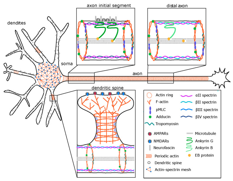
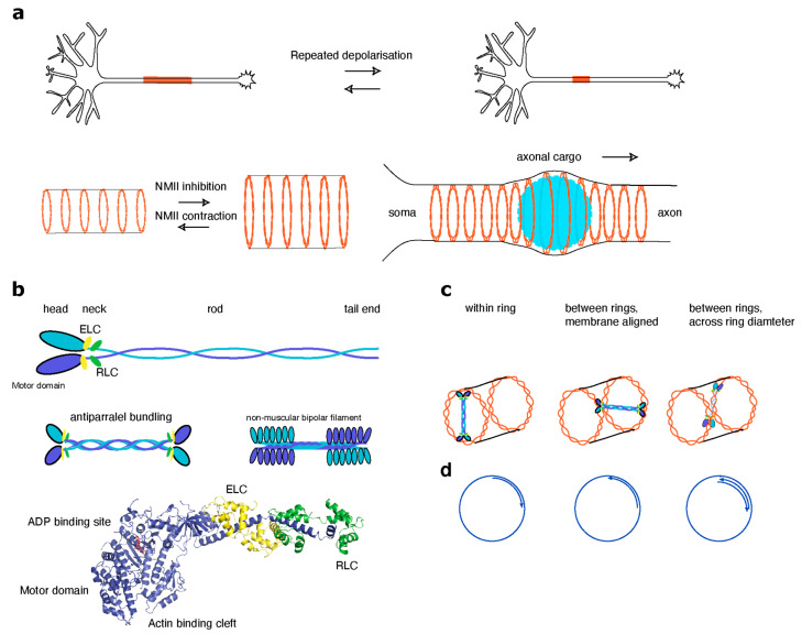
Actin and non-muscle myosins have long been known to play important roles in growth cone steering and neurite outgrowth. More recently, novel functions for non-muscle myosin have been described in axons and dendritic spines. Consequently, possible roles of actomyosin contraction in organizing and maintaining structural properties of dendritic spines, the size and location of axon initial segment and axonal diameter are emerging research topics. In this review, we aim to summarize recent findings involving myosin localization and function in these compartments and to discuss possible roles for actomyosin in their function and the signaling pathways that control them.

Keywords: AIS; actin; calcium signaling; calpain; dendritic spines; myosin; spectrin.


Conflict of interest statement

The authors declare no conflict of interest.

Figures

Figure 1
Figure 1
The organization of the neuronal cytoskeleton. A two-dimensional (2D) actin-spectrin meshwork, similar to those found in other cell types (e.g., erythrocytes), spans the soma of the cell. In contrast, a one-dimensional (1D) periodic membrane cytoskeleton (MPS) is found in axons, in a fraction of dendrites and at the neck of dendritic spines. Top: The MPS consists of actin rings at a periodicity of ~200 nm, interspersed with spectrin tetramers. Each actin ring is formed by two braided actin filaments. The actin rings are further stabilized and regulated by the capping protein adducin and by tropomyosin. The spectrin tetramers are comprised of two αII spectrins and two compartment specific isoforms of β-spectrin. The axon initial segment (AIS) is a stretch of 50–100 µm at the beginning of the axon. The major scaffold in the AIS is AnkyrinG (AnkG), which binds to spectrin βIV and recruits the adhesion molecule neurofascin and ion channels. Phosphorylated myosin light chain (pMLC) is localized to actin rings in the axon. Microtubule bundles are stabilized by plus end binding proteins (EB) along the axon in both the AIS and the distal axon. In the distal axon the MPS is organized by AnkB, which in turn binds to βII-spectrin. AnkB is also arranged periodically, though the pattern is less prominent. Bottom: Dendritic spines are important for compartmentalization of synaptic signaling conferred by glutamate receptors and calcium channels. While the head of the spine contains branched actin filaments, the MPS is prominent in the neck region. Likely consisting of acting rings interspersed by αII and βII-spectrin tetramers. The MPS has also been observed in a sub-fraction of mature dendrites. Here, spectrin tetramers contain the βIII-spectrin isoform.
Figure 2
Figure 2
Organization and function of non-muscle myosin II (NMII). (a, top) Repeated depolarization leads to proximal shortening of the AIS and subsequent extension towards the distal axon. NMII activity is necessary for this process as blebbistatin completely blocks the activity. (a, bottom) NMII activity controls the axonal diameter. Inhibition of NMII by blebbistatin leads to an increase in axon diameter, indicating that NMII holds the membrane-associated cytoskeleton (MSK) under constant tension. In addition, NMII has been shown to be implicated in cargo trafficking along the AIS. The size of large cargo (e.g., autophagosomes, mitochondria, endosomes or lysosomes) can exceed the diameter of the axon. Passage of this large axonal cargo causes a transient radial expansion of the axon followed by constriction, which depend on myosin II activity. (b, top) Schematic organization of NMII. NMII exists as a hexamer that consists of two copies each of elongated heavy chains, two regulatory light chains (RLC) and two essential light chains (ELC) that stabilize the heavy chain structure. The heavy chain is composed of an N-terminal motor domain, a neck domain, which interacts with both light chains, an α-helical rod domain and a C-terminal tail. (b, middle) The hexameric units further bundle both in a parallel and antiparallel manner into bipolar structures that can pull actin filaments together. (b, bottom) Crystal structure of the motor and neck domains of NMII interacting with ELC and RLC. The motor domain contains the actin binding cleft where NMII interacts with actin. Shown in red is ADP bound at the nucleotide binding site. Cycling from ATP to ADP at the nucleotide binding site leads to conformational changes in the actin binding cleft, which modulate the interaction of NMII with actin. (c) Models of a possible spatial relationships between NMII and actin rings. The length of an active two-headed myosin motor complex is around 300 nm, while the distance between actin rings is only 190 nm. (c, left) NMII crosses the diameter of a single actin ring. Alternatively, NMII could cross link two neighboring rings with an angle deviant from 90°, which can be achieved when myosin motors connect neighboring actin rings in a one-dimensional lattice (as spectrin) (c, middle) or across the axonal volume (c, right). (d) Polarity of actin filaments in ring-forming actin braids. Force generated by NMII induces filament sliding within the braid that results in constriction or expansion of the ring. However, as myosin II steps towards the barbed end of an actin filament, the orientation of filaments within a single braid and filament polarity with respect to the neighboring rings are important. The NMII mechanism can work when filaments within a single ring are parallel (d, left) but the adjacent braids have opposite polarity (d, middle) or when two filaments within the same braid have an opposite polarity (c, right).

References

    1. Burnette D.T., Ji L., Schaefer A.W., Medeiros N.A., Danuser G., Forscher P. Myosin II Activity Facilitates Microtubule Bundling in the Neuronal Growth Cone Neck. Dev. Cell. 2008;15:163–169. doi: 10.1016/j.devcel.2008.05.016. - DOI - PMC - PubMed
    1. Kollins K.M., Hy J., Bridgman P.C., Hyang Y.Q., Gallo G. Myosin-II negatiwely regulates minor process extension and the temporal development of neuronal polarity. Dev. Neurobiol. 2009;69:279–298. doi: 10.1002/dneu.20704. - DOI - PMC - PubMed
    1. Yang Q., Zhang X.F., Pollard T.D., Forscher P. Arp2/3 complex-dependent actin networks constrain myosin II function in driving retrograde actin flow. J. Cell Biol. 2012;197:939–956. doi: 10.1083/jcb.201111052. - DOI - PMC - PubMed
    1. Medeiros N.A., Burnette D.T., Forscher P. Myosin II functions in actin-bundle turnover in neuronal growth cones. Nat. Cell Biol. 2006;8:215–226. doi: 10.1038/ncb1367. - DOI - PubMed
    1. Costa A.C.R., Sousa S.C., Pinto-Costa R., Mateus J.C., Lopes C.D.F., Costa A.C.R., Rosa D., Machado D., Pajuelo L., Wang X., et al. The membrane periodic skeleton is an actomyosin network that regulates axonal diameter and conduction. eLife. 2020;9:1–20. doi: 10.7554/eLife.55471. - DOI - PMC - PubMed

Publication types

LinkOut - more resources