. 2020 Sep 1;21(17):6348.
doi: 10.3390/ijms21176348.

The MEK/ERK Module Is Reprogrammed in Remodeling Adult Cardiomyocytes

Affiliations

The MEK/ERK Module Is Reprogrammed in Remodeling Adult Cardiomyocytes

Thomas Kubin et al. Int J Mol Sci. .

Abstract

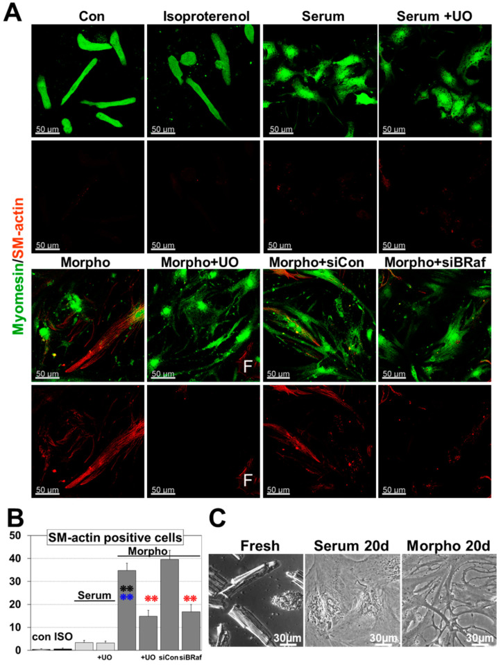
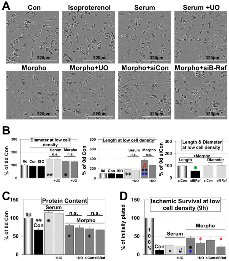
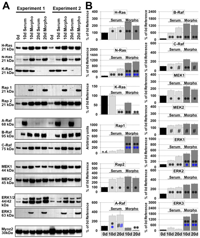
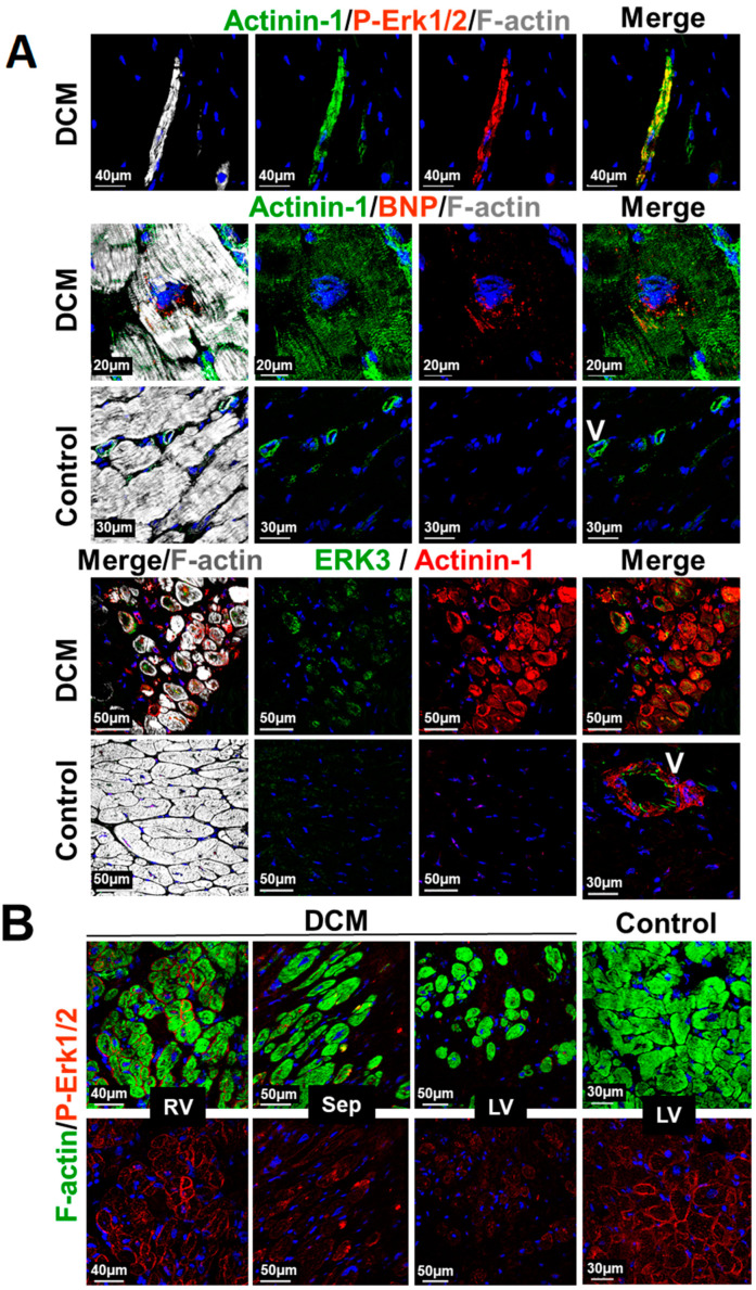
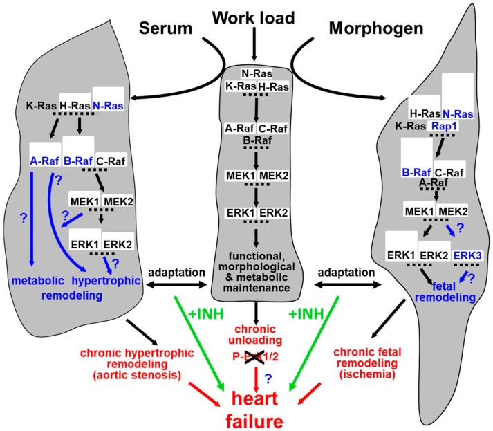
Fetal and hypertrophic remodeling are hallmarks of cardiac restructuring leading chronically to heart failure. Since the Ras/Raf/MEK/ERK cascade (MAPK) is involved in the development of heart failure, we hypothesized, first, that fetal remodeling is different from hypertrophy and, second, that remodeling of the MAPK occurs. To test our hypothesis, we analyzed models of cultured adult rat cardiomyocytes as well as investigated myocytes in the failing human myocardium by western blot and confocal microscopy. Fetal remodeling was induced through endothelial morphogens and monitored by the reexpression of Acta2, Actn1, and Actb. Serum-induced hypertrophy was determined by increased surface size and protein content of cardiomyocytes. Serum and morphogens caused reprogramming of Ras/Raf/MEK/ERK. In both models H-Ras, N-Ras, Rap2, B- and C-Raf, MEK1/2 as well as ERK1/2 increased while K-Ras was downregulated. Atrophy, MAPK-dependent ischemic resistance, loss of A-Raf, and reexpression of Rap1 and Erk3 highlighted fetal remodeling, while A-Raf accumulation marked hypertrophy. The knock-down of B-Raf by siRNA reduced MAPK activation and fetal reprogramming. In conclusion, we demonstrate that fetal and hypertrophic remodeling are independent processes and involve reprogramming of the MAPK.

Keywords: ERK; MEK; Raf; Rap; Ras; dedifferentiation; heart failure; hypertrophy; remodeling; reprogramming.


Conflict of interest statement

The authors declare that they have no competing interests.

Figures

Figure 1
Figure 1
Fetal but not hypertrophic remodeling leads to the reexpression of fetal genes. Primary cultures of adult cardiomyocytes were harvested at the start of the experiment (0 d, reference), after 10 (10 d), and 20 days (20 d) for western blot analysis. Zero-day cultures reflect the protein composition of cardiomyocytes in the normal adult heart. Cultures were stimulated with 5% serum (Serum) and conditioned supernatant of cardiac microvascular endothelial cells containing no growth supplements (Morpho, 80%). (A) Total protein (10 µg) were analyzed for the expression of vinculin, α-tubulin (Tubulin), desmin, skeletal actin (Skel-actin), and the reexpression of α-actinin-1 (Actinin-1), β-actin and α-smooth muscle actin (SM-actin). Muscle actin and pan-actin served as loading controls. Detected molecular sizes are indicated. (B) Quantitative evaluation and statistical analysis of (A). Note that the order of groups is presented differently from (A). Single and double stars indicate p < 0.01 and p < 0.001, respectively. Hash signs indicate p < 0.05. Black signs refer statistically to Con 0 d, blue signs to serum, and the red to all other groups.
Figure 2
Figure 2
Morphogen but not serum induces Ras/Raf/MEK/ERK cascade (MAPK)-dependent remodeling and dedifferentiation. (A) Myomesin and α-smooth muscle actin (SM-actin) fluorescence images after 6-day treatment. SM-actin serves as marker for dedifferentiation/fetal remodeling. Primary cultures of adult cardiomyocytes were untreated or treated with 20 nM isoproterenol (ISO), serum (Serum) and morphogens (Morpho). UO126 (5 µM) was added as an inhibitor of the MAPK pathway (+UO) 1h before experimental start. Note that knock-down of B-Raf (siBRaf) was performed for three days prior to stimulation. Single and double stars indicate p < 0.01 and p < 0.001, respectively. Black signs refer statistically to Con, blue signs to serum, and red compares Morpho (Morpho, Morpho siCon) statistically with and without UO/siB-Raf (+UO, siBRaf). A SM-actin positive fibroblast acts as positive control and is indicated by a white F. (B) Quantitative evaluation of the number of SM-actin positive cells and statistical analysis of (A). (C) Phase-contrast images of freshly isolated cells (Fresh), as well as Serum- (Serum 20 d) and Morpho-treated (Morpho 20 d) cardiomyocytes after 20 d showing long-term effects.
Figure 3
Figure 3
Fetal remodeled but not hypertrophic cardiomyocytes show MAPK dependent ischemic resistance. Primary cultures of adult cardiomyocytes were untreated (Con) or treated with isoproterenol (ISO), serum (Serum), and morphogens (Morpho) for 6 days. Cardiomyocytes were seeded at low density except in (C). UO126 (5 µM) was added (+UO) 1 h before experimental start. Note that cultures were pretreated with siRNA (siCon, siBRaf) three days before experimental start. Single and double stars indicate p < 0.01 and p < 0.001, respectively. Hash signs indicate p < 0.05. Black signs refer statistically to the 0 d group, blue signs to serum, and red compares Morpho (Morpho, Morpho siCon) statistically with and without UO/siB-Raf (+UO, siBRaf). The green star refers to the siCon group. (A) Phase-contrast images of cardiomyocyte cultures. (B) Quantitative and statistical evaluation of cell length and diameter of (A). (C) Total protein content of 6 d-treated cultures at high cell density were compared with cardiomyocytes at experimental start (0 d). (D) Cardiomyocytes were pretreated for 6 days with 0.2 mg/mL albumin (Con), serum, serum + UO126, morphogen, morphogen + UO126. At the end of a 6-day culture period, all cells were counted (reference value 100%) and then kept in glucose-free PBS at 1% O2. After 9 h of ischemia, cardiomyocytes were fixed in 4% paraformaldehyde and recounted.
Figure 4
Figure 4
Knock-down of B-Raf and treatment with UO126 (+UO) abrogates MAPK cascade activation and fetal remodeling. Isolated cardiomyocytes (n = 4) were allowed to recover for 1 day (Con 0 d) and then treated for 10 min (10 min) or 6 days (6 d) for western blot analysis. Cultures were treated with basic medium (Con), with serum (Serum), or with morphogens (Morpho) as indicated. MEK1/2 inhibitor UO126 (UO, 5 µM) was added 1 h before the experimental start as indicated. Quantitative evaluation and statistical analysis of western blots (WB) are shown. Single star and hash sign indicate p < 0.01 and p < 0.05, respectively. Black signs refer statistically to Con. Red signs compare Morpho (Morpho, Morpho siCon) statistically with and without UO/siB-Raf (+UO, siBRaf). (A) WB analysis of ERK1/2 (P-ERK1/2) activation and dedifferentiation (SM-actin) in the absence or presence of UO (+UO). (B) WB analysis of P-ERK1/2 of untreated cardiomyocytes (cultured in basic medium) at 0 d and after 6 d (n = 6). (C) After a three-day pretreatment period with siRNA, cardiomyocytes were cultured in basic medium or stimulated with Serum and Morpho for WB of α-actinin-1 (Actinin-1) and α-smooth muscle actin (SM-actin) for six days. In order to determine MEK1/2 and ERK1/2 phosphorylation (P-MEK1/2, P-ERK1/2) cultures were made quiescent after six days for one day in basic medium and then treated as indicated for 10 min.
Figure 5
Figure 5
MAPK remodeling in dedifferentiating cardiomyocytes is distinct from hypertrophic myocytes. Primary cultures of adult cardiomyocytes were harvested at the experimental start (0 d, reference), and after 10 (10 d), and 20 days (20 d) for western blot analysis. Cultures were stimulated with 5% serum (Serum) and conditioned supernatant (Morpho, 80%) of cardiac microvascular endothelial cells containing no growth supplements. Single and double stars indicate p < 0.01 and p < 0.001, respectively. Black stars refer statistically to Con, blue stars to serum. Hash signs indicate p < 0.05. For A-Raf blue symbols refer statistically to morphogens. (A) Total protein (10 µg) was analyzed for the expression of components of the Ras/Raf/MEK/ERK pathway, as well as the Ras-related proteins Rap1 and Rap2. MYOZ is Calsarcin-1 and serves as a loading control. Note that we detected Erk3 as the 63 kDa form but not as full length 90 kDa protein. Detected molecular sizes are indicated. (B) Quantitative evaluation and statistical analysis of (A). Note that the order of groups is presented differently from (A).
Figure 6
Figure 6
Structural and MAPK remodeling of cardiomyocytes in the myocardium of patients with dilated cardiomyopathy. (A) Confocal images of a transplanted patient with end-stage dilated cardiomyopathy (DCM). Upper images show an α-actinin-1 (Actinin-1)- and P-Erk-1/2-positive cell isolated from other cardiomyocytes in a fibrotic area of the myocardium. P-ERK1/2 indicates that the MAPK is still intact. Middle images depict α-actinin-1 positive cardiomyocyte expressing BNP. Lower images show cardiomyocytes reexpressing α-actinin-1 as well as Erk3. Controls show neither ERK3 nor actinin-1 expression. V indicates a vessel and serves as positive control for actinin-1. (B) Confocal fluorescence images of a transplanted 12-year-old patient with left ventricular assist device. Note the P-ERK1/2-decreasing gradient from the right ventricle (RV) over the septum (Sep) to the left ventricle (LV). LV control serves an age-matched patient with Tetralogy of Fallot.
Figure 7
Figure 7
Remodeling of the MAPK pathway in the hypothetical context of cardiac adaptation and failure. The myocardium has a certain capacity to adapt to increased load either by hypertrophy (e.g., evoked though aortic stenosis) or by fetal remodeling (e.g., evoked through ischemia). However, when cardiac demands are not met and become chronic, heart failure may develop. Cardiomyocyte remodeling does not only result in structural reorganization but also involves reprogramming of the MAPK. A third road to heart failure might be produced by cardiac unloading due to the absence of appropriate stimuli rendering cardiomyocytes unfunctional. The interference by MAPK inhibitors (+INH) might disturb adaptive processes and enhance the development of heart failure.

References

    1. Swynghedauw B. Molecular mechanisms of myocardial remodeling. Am. J. Physiol. 1999;79:215–262. doi: 10.1152/physrev.1999.79.1.215. - DOI - PubMed
    1. Cohn J.N., Ferrari R., Sharpe N. Cardiac Remodeling—Concepts and Clinical Implications: A Consensus Paper From an International Forum on Cardiac Remodeling. J. Am. Coll. Cardiol. 2000;35:569–582. doi: 10.1016/S0735-1097(99)00630-0. - DOI - PubMed
    1. Hein S., Arnon E., Kostin S., Schonburg M., Elsasser A., Polyakova V., Bauer E.P., Klovekorn W.-P., Schaper J. Progression From Compensated Hypertrophy to Failure in the Pressure-Overloaded Human Heart: Structural Deterioration and Compensatory Mechanisms. Circulation. 2003;107:984–991. doi: 10.1161/01.CIR.0000051865.66123.B7. - DOI - PubMed
    1. Hoshijima M., Chien K.R. Mixed signals in heart failure. J. Clin. Investig. 2002;109:849–855. doi: 10.1172/JCI0215380. - DOI - PMC - PubMed
    1. Banks M., Crowell K., Proctor A., Jensen B.C. Cardiovascular Effects of the MEK Inhibitor, Trametinib: A Case Report, Literature Review, and Consideration of Mechanism. Cardiovasc. Toxicol. 2017;17:487–493. doi: 10.1007/s12012-017-9425-z. - DOI - PMC - PubMed

MeSH terms

Substances