. 2020 Sep 1;12(9):2483.
doi: 10.3390/cancers12092483.

Lenalidomide in Combination with Arsenic Trioxide: an Effective Therapy for Primary Effusion Lymphoma

Affiliations

Lenalidomide in Combination with Arsenic Trioxide: an Effective Therapy for Primary Effusion Lymphoma

Sara Moodad et al. Cancers (Basel). .

Abstract

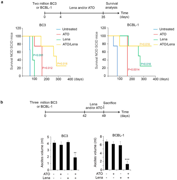
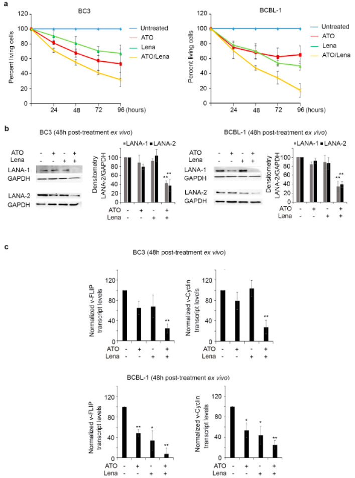
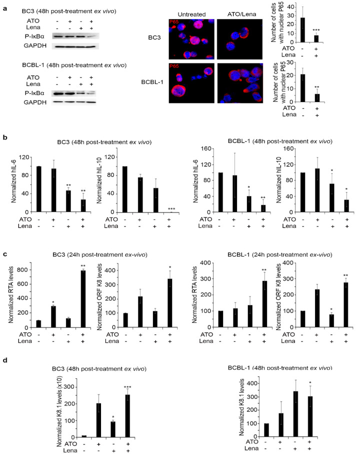
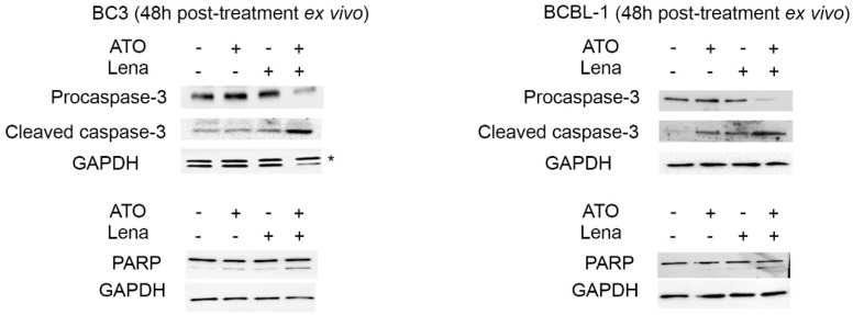
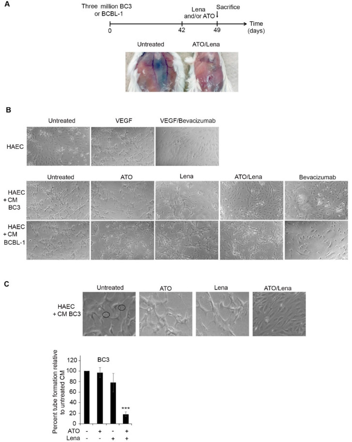
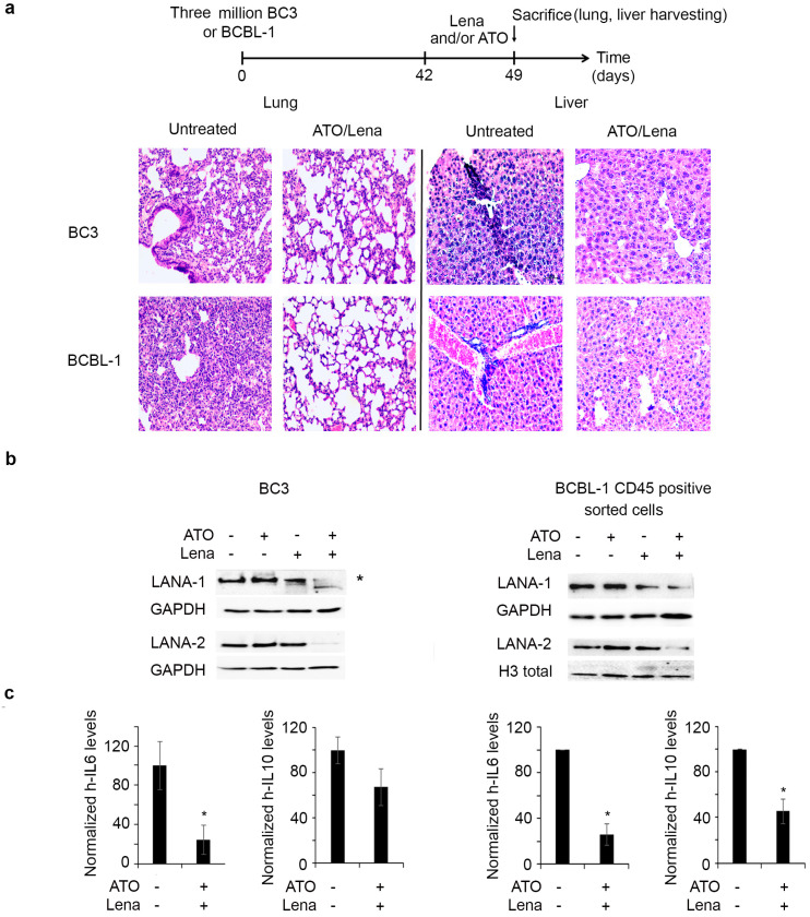
Primary effusion lymphoma (PEL) is a rare aggressive subset of non-Hodgkin B cell lymphoma. PEL is secondary to Kaposi sarcoma herpes virus (KSHV) and predominantly develops in serous cavities. Conventional chemotherapy remains the treatment of choice for PEL and yields high response rates with no significant comorbidities. Yet, chemotherapy often fails in achieving or maintaining long-term remission. Lenalidomide (Lena), an immunomodulatory drug, displayed some efficacy in the treatment of PEL. On the other hand, arsenic trioxide (ATO) in combination with other agents effectively treated a number of blood malignancies, including PEL. In this study, we present evidence that the combination of ATO/Lena significantly enhanced survival of PEL mice, decreased the volume of exacerbated ascites in the peritoneum, and reduced tumor infiltration in organs of treated animals. In ex vivo treated PEL cells, ATO/Lena decreased the proliferation and downregulated the expression of KSHV latent viral proteins. This was associated with decreased NF-κB activation, resulting in reactivation of viral replication, downregulation of interleukin-6 (IL-6) and IL-10, inhibition of vascular endothelial growth factor, and apoptosis. Our results elucidate the mechanism of action of ATO/Lena and present it as a promising targeted therapeutic modality in PEL management, which warrants further clinical investigation.

Keywords: HHV-8; LANA; immunomodulatory drugs; latent cycle; lymphoma; lytic cycle.


Conflict of interest statement

The authors declare no conflict of interest.

Figures

Figure 1
Figure 1
Arsenic trioxide/Lenalidomide (ATO/Lena) enhanced survival and decreased ascites volume in NOD/SCID primary effusion lymphoma (PEL) mice. (a) Kaplan-Meier graphs of overall survival curves of BC-3 (left) and BCBL-1 (right) NOD/SCID mice. Mice (n = 4 per condition) were injected with 2 million BC-3 or BCBL-1 cells. ATO, Lena, or their combination were administered from day 4 until day 35 post-injection of PEL cells. (b) Ascites volume from BC-3 (left) or BCBL-1 (right). PEL mice were allowed to develop ascites for 6 weeks then were treated daily with ATO, Lena, or their combination for one week before sacrifice. (**) indicates p < 0.01; and (***) indicates p < 0.001.
Figure 2
Figure 2
ATO/Lena inhibited proliferation and downregulated Kaposi sarcoma herpes virus (KSHV) latent transcripts and proteins in ex vivo treated ascites-derived BC-3 and BCBL-1 cells. (a) Cell proliferation of ascites-derived BC-3 (left) or BCBL-1 cells (right) following ex vivo treatment with ATO and/or Lena for 24, 48, 72, and 96 h. Results are presented as percent of control, plotted as mean ± SD, and represent an average of three independent experiments. (b) Immunoblot analysis of KSHV latent proteins LANA-1 and LANA-2 in ascites-derived BC-3 (left) or BCBL-1(right) cells treated ex vivo for 48 h with ATO, Lena, or their combination. Densitometry histograms represent an average of 3 independent experiments. Uncropped blots of Figure 2b are shown in Figure S5 (c) Real-time quantitative PCR analysis of transcript levels of KSHV latent genes v-FLIP and v-Cyclin in ascites-derived BC-3 (left) or BCBL-1(right), 48 h post treatment with ATO, Lena, or the ATO/Lena combination. Results represent the average of 3 independent experiments. (*) indicates p < 0.05; (**) indicates p < 0.01.
Figure 3
Figure 3
ATO/Lena inhibited NF-κB activation and increased KSHV reactivation in ex vivo treated ascites-derived BC-3 and BCBL-1 cells. (a) Western blot analysis of p-IκBα in ATO/Lena ex vivo treated ascites-derived BC-3 and BCBL-1 cells 48 h post treatment. Confocal microscopy analysis of p65 nuclear translocation in ascites-derived BC-3 and BCBL-1 after ex vivo treatment with ATO/Lena for 48 h. p65 was stained with anti-p65 antibody (red) and nuclei were stained by Hoechst stain (blue). Images represent z-sections. Histograms represented number of cells with nuclear p65 translocation. Uncropped blots of Figure 3a are shown in Figure S5. (b) Real-time quantitative PCR showing transcript levels of human IL-6 and IL-10 cytokines in ascites-derived BC-3 (left) and BCBL-1 (right) cells 48 h following ex vivo treatment with ATO and/or Lena. (c,d) Real-time quantitative PCR analysis of transcript levels of KSHV early-lytic genes (RTA, ORFK8) (c) or late lytic gene (K8.1) (d) in ascites-derived BC-3 (left) and BCBL-1 (right) cells after ex vivo treatment with ATO and/or Lena for 24 (c) or 48 h (d) as indicated. Results represent the average of 3 independent experiments. (*) indicates p < 0.05; (**) indicates p < 0.01; and (***) indicates p < 0.001.
Figure 4
Figure 4
The ATO/Lena combination induced apoptosis in ex vivo treated ascites-derived BC-3 and BCBL-1 cells. Western blot analysis of PARP, procaspase-3, and cleaved caspase-3 levels in ascites-derived BC-3 (left) and BCBL-1 (right) cells 48 h post treatment as indicated. Uncropped blots are shown in Figure S5.
Figure 5
Figure 5
ATO/Lena decreased VEGF-induced tubal formation potential of ascites-derived BC-3 but not BCBL-1 cells. (A) Mice peritoneum vascularization before and after one-week treatment with ATO/Lena in BC-3 PEL mice. (B) Light microscopy images of capillary-like tube formations in HAEC cells following incubation with supernatants from ex vivo treated ascites-derived BC-3 or BCBL-1 cells. (C) Tubal formation analysis method where nodes with 3 or more branches were counted and compared. At least 5 images from each condition were counted. Data was reported as a histogram of the percentage of tube formation relative to untreated ascites-derived BC-3 cells. Data represent an average of 3 independent experiments. (***) indicates p < 0.001.
Figure 6
Figure 6
ATO/Lena decreased organ infiltration and downregulated latent KSHV proteins in vivo. (a) Experimental design: mice injected with BC-3 or BCBL-1 were allowed to develop ascites for 6 weeks, were treated with ATO and/or Lena for one week, then were sacrificed. Histopathology sections of lung and spleens from ATO/Lena treated or untreated BC-3 and BCBL-1 PEL mice. (b). Immunoblot analysis of LANA-1 and LANA-2 proteins in whole ascites (left) or CD45+ sorted ascites (right) derived from treated or untreated BC-3 and BCBL-1 mice respectively. Uncropped blots of Figure 6b are shown in Figure S5. (c) Real-time quantitative PCR analysis of cellular IL-6 and IL-10 transcript levels in lungs of ATO/Lena treated or untreated BC-3 and BCBL-1 mice as indicated. Transcript levels were normalized to GAPDH internal levels. (*) indicates p < 0.05

Similar articles

Cited by

References

    1. Katano H. Pathological Features of Kaposi’s Sarcoma-Associated Herpesvirus Infection. In: Kawaguchi Y., Mori Y., Kimura H., editors. Human Herpesviruses. Springer; Singapore: 2018. pp. 357–376. - PubMed
    1. Shimada K., Hayakawa F., Kiyoi H. Biology and management of primary effusion lymphoma. Blood. 2018;132:1879–1888. doi: 10.1182/blood-2018-03-791426. - DOI - PubMed
    1. Chang Y., Cesarman E., Pessin M., Lee F., Culpepper J., Knowles D., Moore P. Identification of herpesvirus-like DNA sequences in AIDS-associated Kaposi’s sarcoma. Science. 1994;266:1865–1869. doi: 10.1126/science.7997879. - DOI - PubMed
    1. Cesarman E., Chang Y., Moore P.S., Said J.W., Knowles D.M. Kaposi’s Sarcoma–Associated Herpesvirus-Like DNA Sequences in AIDS-Related Body-Cavity–Based Lymphomas. N. Engl. J. Med. 1995;332:1186–1191. doi: 10.1056/NEJM199505043321802. - DOI - PubMed
    1. Minhas V., Wood C. Epidemiology and Transmission of Kaposi’s Sarcoma-Associated Herpesvirus. Viruses. 2014;6:4178–4194. doi: 10.3390/v6114178. - DOI - PMC - PubMed

LinkOut - more resources