. 2020 Sep 1;21(17):6355.
doi: 10.3390/ijms21176355.

Assessing Lysosomal Disorders in the NGS Era: Identification of Novel Rare Variants

Affiliations

Assessing Lysosomal Disorders in the NGS Era: Identification of Novel Rare Variants

Marisa Encarnação et al. Int J Mol Sci. .

Abstract

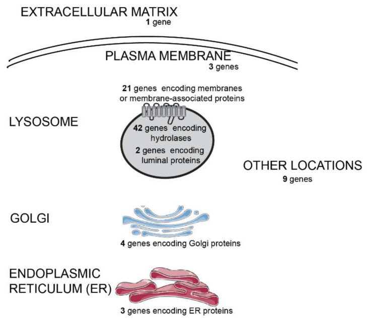
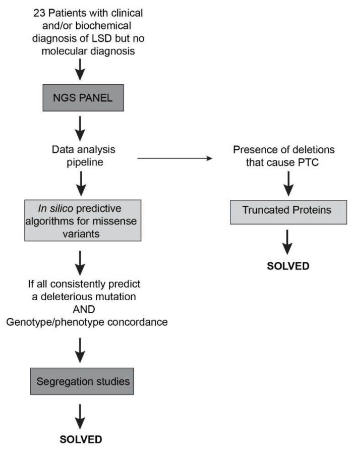
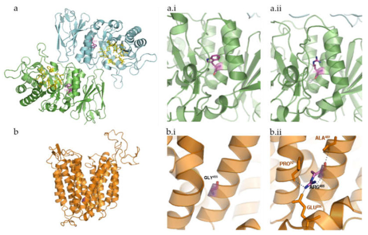
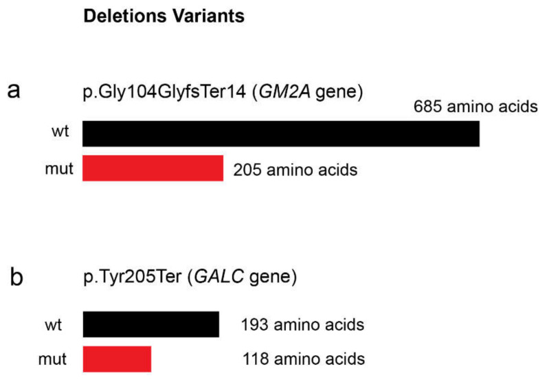
Lysosomal storage diseases (LSDs) are a heterogeneous group of genetic disorders with variable degrees of severity and a broad phenotypic spectrum, which may overlap with a number of other conditions. While individually rare, as a group LSDs affect a significant number of patients, placing an important burden on affected individuals and their families but also on national health care systems worldwide. Here, we present our results on the use of an in-house customized next-generation sequencing (NGS) panel of genes related to lysosome function as a first-line molecular test for the diagnosis of LSDs. Ultimately, our goal is to provide a fast and effective tool to screen for virtually all LSDs in a single run, thus contributing to decrease the diagnostic odyssey, accelerating the time to diagnosis. Our study enrolled a group of 23 patients with variable degrees of clinical and/or biochemical suspicion of LSD. Briefly, NGS analysis data workflow, followed by segregation analysis allowed the characterization of approximately 41% of the analyzed patients and the identification of 10 different pathogenic variants, underlying nine LSDs. Importantly, four of those variants were novel, and, when applicable, their effect over protein structure was evaluated through in silico analysis. One of the novel pathogenic variants was identified in the GM2A gene, which is associated with an ultra-rare (or misdiagnosed) LSD, the AB variant of GM2 Gangliosidosis. Overall, this case series highlights not only the major advantages of NGS-based diagnostic approaches but also, to some extent, its limitations ultimately promoting a reflection on the role of targeted panels as a primary tool for the prompt characterization of LSD patients.

Keywords: CLN7; GM2 Gangliosidosis; GM2A gene; bioinformatics analysis; diagnostics odyssey; lysosomal storage diseases (LSDs); molecular genetic testing (MGT); next-generation sequencing (NGS).


Conflict of interest statement

The authors declare no conflict of interest.

Figures

Figure 1
Figure 1
Subcellular distribution of the different gene products whose coding sequences were included in our next generation sequencing (NGS) panel designed to diagnose lysosomal storage diseases (LSDs) and related disorders. The panel is composed by 85 genes, as depicted: 65 genes encoding lysosomal proteins and 20 genes coding for non-lysosomal proteins. Some genes associated to diseases resembling LDSs or genes encoding proteins associated with lysosome-related organelles (LROs) were also covered, in such a way that, from a pathophysiological point of view, the panel includes 54 LSD-related genes, 13 which are disease-causing but non-LSD-related, and 18 not previously associated to disease. The non-LSD-disease-causing genes include three involved in LRO disorders and 10 whose deficiency is known to underlie disorders with overlapping phenotypes.
Figure 2
Figure 2
Workflow of the study of the patients and respective molecular characterization The NGS-targeted gene panel allowed the identification of four novel and six previously identified pathogenic variants in a group of 23 patients. PTC = premature termination codon.
Figure 3
Figure 3
Three-dimensional structure representation of α-galactosidase (GLA) and major facilitator superfamily domain containing 8 (MFSD8) using PyMol. β-strands, helices, and coils indicate the secondary structure elements that form the scaffold for the interacting residues. Relevant side chains that interact with the residues of interest are depicted and labelled. (a) Human GLA dimer, shown in ribbon representation (Protein Data Bank accession code = 1R46). Each monomer of the homodimer contains two domains, α(β/α)8 barrel containing the active site plus a C-terminal antiparallel β domain. Active site residues are shown in yellow (Trp47, Asp92, Asp93, Tyr134, Cys142, Lys168, Asp170, Glu203, Leu206, Tyr207, Arg227, Asp231, Asp266, Met267). Relevant side chains that interact with the residue of interest are depicted and labelled. α-galactosidase A dimer, with the active site residues and the affected amino acid highlighted; (a.i) wild-type Trp287; (a.ii)—mutated Arg287; (b) in silico design of MFSD8 three-dimensional structure based on sequence analysis and subsequent prediction of the secondary structure elements it encodes. The three-dimensional structure depicted does not take into consideration the fact that this is a multi-pass membrane protein. Instead, it refers to the mature protein MFSD8 as it would coil based solely on the intra-molecular interactions. Still, a comparison between this prediction and that of the transmembrane domains further validates the theoretical assumption that MFSD8 contains 12 transmembrane domains, which are formed by α-helices. (b.i) Wild-type Gly455. (b.ii) Mutated Arg455.
Figure 4
Figure 4
Prevision of the effect of the novel identified deletions ((a) c.312del in GM2A gene and (b) c.613_617del in GALC gene) over protein size. wt = non mutated protein; mut = mutated protein.

References

    1. Schultz M.L., Tecedor L., Chang M., Davidson B.L. Clarifying lysosomal storage diseases. Trends Neurosci. 2011;34:401–410. doi: 10.1016/j.tins.2011.05.006. - DOI - PMC - PubMed
    1. Futerman A.H., Van Meer G. The cell biology of lysosomal storage disorders. Nat. Rev. Mol. Cell Biol. 2004;5:554–565. doi: 10.1038/nrm1423. - DOI - PubMed
    1. Platt F.M., d’Azzo A., Davidson B.L., Neufeld E.F., Tifft C.J. Lysosomal storage diseases. Nat. Rev. Dis. Prim. 2018 doi: 10.1038/s41572-018-0025-4. - DOI - PubMed
    1. Winchester B. Lysosomal diseases: Diagnostic update. J. Inherit. Metab. Dis. 2014;37:599–608. doi: 10.1007/s10545-014-9710-y. - DOI - PubMed
    1. Filocamo M., Morrone A. Lysosomal storage disorders: Molecular basis and laboratory testing. Hum. Genom. 2011 doi: 10.1186/1479-7364-5-3-156. - DOI - PMC - PubMed

Substances