Case Reports
. 2020 Sep 3;20(1):195.
doi: 10.1186/s12893-020-00857-x.

A case report of severe degenerative lumbar scoliosis associated with windswept lower limb deformity

Affiliations
Case Reports

A case report of severe degenerative lumbar scoliosis associated with windswept lower limb deformity

Xi Yang et al. BMC Surg. .

Abstract

Background: The windswept lower limb deformity describes valgus deformity in one leg with varus deformity in the other. It is mostly seen in young children with metabolic bone diseases (such as rickets) and may lead to leg length discrepancy (LLD) and Degenerative scoliosis (DS) in older age. To the best of our knowledge, there was no report of the spinal surgery in patient with severe DS associated with windswept deformity. The objective of this study is to report the unique case of a 60-year-old woman with severe degenerative scoliosis (DS) associated with windswept deformity caused by rickets who underwent a posterior correction and fusion surgery in spine.

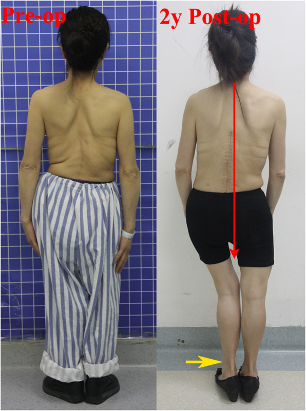
Case presentation: The patient was diagnosed as rickets windswept lower limb deformity for 50 years but never went through routine treatment. Then, she performed lumbar scoliosis for more than 20 years and suffered from severe back pain for 4 years. After overall clinical evaluation and radiographic measures, we performed a posterior surgical correction and fusion from T9-L5. With this surgery, the main thoracolumbar curve Cobb angle corrected from 72.5° to 21.0°, the coronal balance from 0 cm to 2.0 cm while the sagittal vertical axis (SVA) from 1.5 cm to - 1.0 cm. At 2 years postoperative follow-up, her back pain has almost completely relieved with a satisfied fixation and bone fusion showed on CT scans. However, a coronal imbalance was found with C7-CSVLdistance equal to 4.0 cm. This coronal imbalance was highly correlated to the untreated LLD and pelvic obliquity, and should be improved by standing posture or shoe lifts.

Conclusions: For such patient, the pure spinal correction and fusion surgery, in spite of lower limbs deformity, can achieve good relieve of back pain symptom, however may accompany by the complication of coronal imbalance due to the unimproved pelvic obliquity and LLD. However, longer follow-up is necessary to observe the long-term outcome of this patient's postoperative coronal imbalance.

Keywords: Degenerative scoliosis; Leg length discrepancy; Pelvic obliquity; Rickets; Windswept lower limb deformity.


Conflict of interest statement

The authors declare that they have no competing interests.

Figures

Fig. 1
Fig. 1
Standing whole length X-ray films before operation. The A-P film shows her main thoracolumbar (TL) curve Cobb is 72.5° (between T10-L3) while lumbosacral (LS) semi-curve Cobb (between L3-S1) is 44.9°. The length of left leg is 66.5 cm that is 2.5 cm longer than right leg (64.1 cm). The leg length discrepancy leads to pelvic obliquity with the pelvic tilt (Pt) angle is 7.1° (left higher than right). And L5 tile (L5t) angle is 21.9°. The coronal balance measured as distance between C7 centre point to CSVL is 0 at this time. The mechanical axis (brown dashed) shows the valgus leg in left side and the varus leg in right side. The bowed long bone deformities are shown in both legs. The lateral film (right side) shows her Pelvic incidence (PI) is 41°, sacral slope (SS) 37.3°, lumbar lordosis (LL) 46.5°, thoracic kyphosis (TK) 15.2° and sagittal vertical axis (SVA) 1.5 cm. Those parameters show her sagittal balance is good
Fig. 2
Fig. 2
Standing whole length X-ray films at 2 years after operation. The A-P film (left side) shows her main thoracolumbar (TL) Cobb is 19.5° while lumbosacral (LS) Cobb is 16.7°. Pelvic tilt (Pt) angle is 7.0° while L5 tile (L5t) angle is 10.8°. Coronal balance (CB) means the distance between C7 centre point to CSVL line (yellow plumb dashed). Its value is 4.0 cm that means radiographic coronal imbalance. However, when we draw the modified CSVL (red dashed) based on the connect line of bilateral iliac highest point (white solid line), measure the distance between C7 centre point to modified CSVL and find the modified coronal balance (modified CB) is 0.6 cm (< 2 cm) which means coronal balance. The lateral film (right side) shows her sagittal balance is well maintained at this time
Fig. 3
Fig. 3
Preoperative (left) and 2 years postoperative appearance (right). Interesting phenomenon is the patient spontaneously moves her left foot insider (yellow arrow) to reduce the practical leg length discrepancy and then get better trunk balance (red line shows the line of gravity) after operation

Similar articles

References

    1. Hong JY, Suh SW, Modi HN, et al. The prevalence and radiological findings in 1347 elderly patients with scoliosis. J Bone Joint Surg Br. 2010;92(7):980–983. doi: 10.1302/0301-620X.92B7.23331. - DOI - PubMed
    1. Ploumis A, Transfledt EE, Denis F. Degenerative lumbar scoliosis associated with spinal stenosis. Spine J. 2007;7(4):428–436. doi: 10.1016/j.spinee.2006.07.015. - DOI - PubMed
    1. Jimbo S, Kobayashi T, Aono K, et al. Epidemiology of degenerative lumbar scoliosis: a community based cohort study. Spine (Phila Pa 1976) 2012;37(20):1763–1770. doi: 10.1097/BRS.0b013e3182575eaa. - DOI - PubMed
    1. Marty-Poumarat C, Scattin L, Marpeau M, et al. Natural history of progressive adult scoliosis. Spine (Phila Pa 1976) 2007;32(11):1227–1234. doi: 10.1097/01.brs.0000263328.89135.a6. - DOI - PubMed
    1. Raczkowski JW, Daniszewska B, Zolynski K. Functional scoliosis caused by leg length discrepancy. Arch Med Sci. 2010;6(3):393–398. doi: 10.5114/aoms.2010.14262. - DOI - PMC - PubMed

Publication types