A novel arthroscopically assisted reduction technique for three patterns of posterolateral tibial plateau fractures
- PMID: 32883325
- PMCID: PMC7469271
- DOI: 10.1186/s13018-020-01901-5
A novel arthroscopically assisted reduction technique for three patterns of posterolateral tibial plateau fractures
Abstract
Background: Posterolateral tibial plateau fractures (PTPF) remain a challenge for orthopedics surgeons because the special anatomical structures of the posterolateral corner of knee joint including the fibular head, the lateral collateral ligament, and the peroneal nerve, which impedes the exposure of the fracture fragments and need irregular implants to get a stable fixation. The purpose of present study was to introduce a new articular fracture fragments restoration technique for three patterns of PTPF and investigate the relationship between associated soft injuries and fracture patterns.
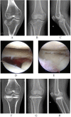
Methods: From May 2016 to April 2018, 31 patients with PTPF who had undertaken arthroscopically assisted reduction and fixation (AARF) were enrolled in present study. Demographic data, pre-operation, and post-operation X plan films, three-dimensional computed tomography (CT) scans and magnetic resonance imaging (MRI) were reviewed. Present samples were divided into three patterns with lateral inclination (LI), posterior inclination (PI), and parallel compression (PC) according to the orientation of the articular fragment inclination. Rasmussen anatomical score was used to assess the radiological results. Rasmussen functional score, Hospital for Special Surgery knee-rating Score (HSS), and range of motion (ROM) of the knee joint at the final follow-up were measured to evaluate the clinical outcomes.
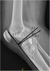
Results: In this series, the post-operation tibial plateau angle (TPA) was 9.7° ± 3.5°(range 4.0°-15.8°) and the Rasmussen anatomical score was 17.7 ± 0.7(range 16-18); clinical outcomes showed that the HSS score was 92.7 ± 21.8 (range 90-96) and the Rasmussen functional score was 27.9 ± 1.0 (range 26-30). Of all the patients, the anterior cruciate ligament (ACL) injuries including the ACL tibial attachment ruptures occurred in 16 patients (51.6%), meniscus lesions happened in 19 patients (61.3%), medial collateral ligament (MCL) injuries were founded in 13 patients (41.9%). The number of ACL injuries including the ACL tibial attachment ruptures in the PI fracture pattern (12 cases) is significantly higher than LI (2 cases) and PC (2 cases) fracture pattern (p < 0.05).
Conclusion: Profound understanding the different patterns of PTPF and using our reduction technique will facilitate to restore the main articular fracture fragments. The PI fracture patterns have a significant high incidence of the ACL ruptures.
Level of evidence: Therapeutic study, Level IV.
Keywords: Arthroscopically assisted reduction and fixation; Fracture patterns; Posterolateral tibial plateau fracture; Restoration; Soft tissue injury.
Conflict of interest statement
The authors declare that they have no competing interests.
Figures


Similar articles
-
Effectiveness of bone grafting versus cannulated screw fixation in the treatment of posterolateral tibial plateau compression fractures with concomitant ACL injury: a comparative study.J Orthop Surg Res. 2024 Jan 17;19(1):75. doi: 10.1186/s13018-023-04516-8. J Orthop Surg Res. 2024. PMID: 38233925 Free PMC article.
-
Clinical results after surgical treatment of posterolateral tibial plateau fractures ("apple bite fracture") in combination with ACL injuries.Eur J Trauma Emerg Surg. 2020 Dec;46(6):1239-1248. doi: 10.1007/s00068-020-01509-8. Epub 2020 Sep 26. Eur J Trauma Emerg Surg. 2020. PMID: 32980883
-
Correlation of parameters on preoperative CT images with intra-articular soft-tissue injuries in acute tibial plateau fractures: A review of 132 patients receiving ARIF.Injury. 2017 Mar;48(3):745-750. doi: 10.1016/j.injury.2017.01.043. Epub 2017 Jan 30. Injury. 2017. PMID: 28190582
-
Arthroscopy-assisted surgery: The management of posterolateral tibial plateau depression fracture accompanying ligament injury: A case series and review of the literature.J Orthop Surg (Hong Kong). 2020 Jan-Apr;28(1):2309499019891208. doi: 10.1177/2309499019891208. J Orthop Surg (Hong Kong). 2020. PMID: 31876260 Review.
-
Open reduction and internal fixation in a one-stage anterior cruciate ligament reconstruction surgery for the treatment of tibial plateau fractures: A case report and literature review.Injury. 2018 Jun;49(6):1215-1219. doi: 10.1016/j.injury.2018.03.015. Epub 2018 Mar 15. Injury. 2018. PMID: 29655591 Review.
Cited by
-
Comparison of outcomes of ORIF versus bidirectional tractor and arthroscopically assisted CRIF in the treatment of lateral tibial plateau fractures: a retrospective cohort study.J Orthop Surg Res. 2021 May 3;16(1):289. doi: 10.1186/s13018-021-02447-w. J Orthop Surg Res. 2021. PMID: 33941204 Free PMC article.
-
The treatment of posterolateral tibial plateau fracture with a newly designed anatomical plate via the trans-supra-fibular head approach: preliminary outcomes.BMC Musculoskelet Disord. 2021 Sep 18;22(1):804. doi: 10.1186/s12891-021-04684-w. BMC Musculoskelet Disord. 2021. PMID: 34537030 Free PMC article.
-
Surgical exposure to posterolateral quadrant tibial plateau fractures: an anatomic comparison of posterolateral and posteromedial approaches.J Orthop Surg Res. 2022 Jul 15;17(1):346. doi: 10.1186/s13018-022-03236-9. J Orthop Surg Res. 2022. PMID: 35841047 Free PMC article.
-
Optimizing Surgical Management of Tibial Plateau Fractures: A Comparative Study of Minimally Invasive Versus Open Reduction Techniques.Cureus. 2024 May 11;16(5):e60078. doi: 10.7759/cureus.60078. eCollection 2024 May. Cureus. 2024. PMID: 38860085 Free PMC article.
-
Effectiveness of bone grafting versus cannulated screw fixation in the treatment of posterolateral tibial plateau compression fractures with concomitant ACL injury: a comparative study.J Orthop Surg Res. 2024 Jan 17;19(1):75. doi: 10.1186/s13018-023-04516-8. J Orthop Surg Res. 2024. PMID: 38233925 Free PMC article.
References
MeSH terms
Grants and funding
LinkOut - more resources
Full Text Sources
Medical
Research Materials
Miscellaneous

