Review
. 2020 Sep 8;76(10):1226-1243.
doi: 10.1016/j.jacc.2020.06.076.

Coronary Computed Tomography Angiography From Clinical Uses to Emerging Technologies: JACC State-of-the-Art Review

Affiliations
Review

Coronary Computed Tomography Angiography From Clinical Uses to Emerging Technologies: JACC State-of-the-Art Review

Khaled M Abdelrahman et al. J Am Coll Cardiol. .

Abstract

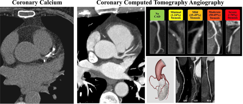
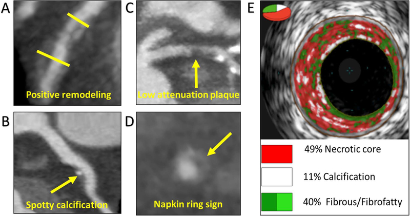
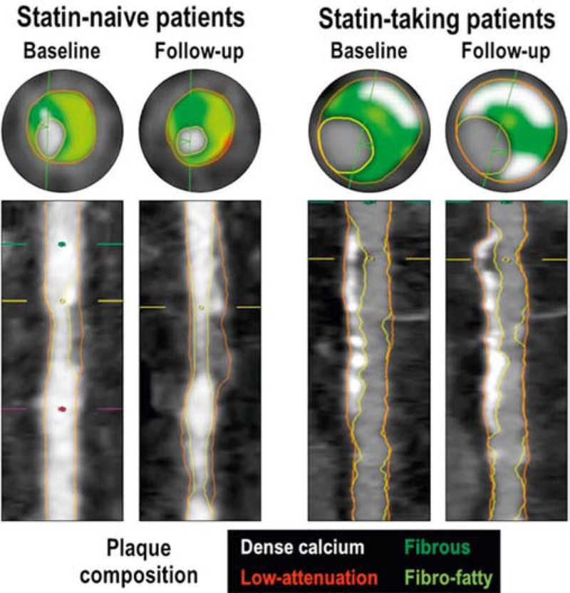
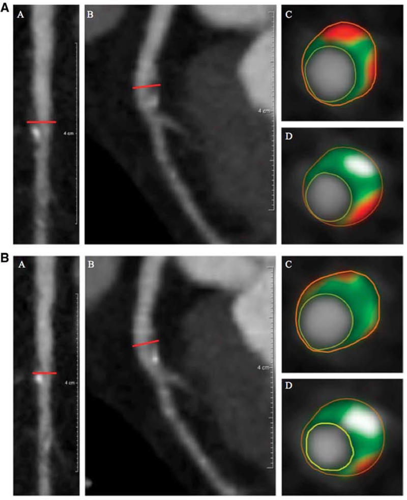
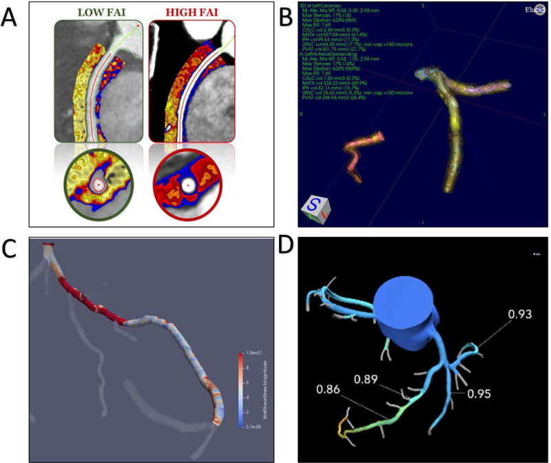
Evaluation of coronary artery disease (CAD) using coronary computed tomography angiography (CCTA) has seen a paradigm shift in the last decade. Evidence increasingly supports the clinical utility of CCTA across various stages of CAD, from the detection of early subclinical disease to the assessment of acute chest pain. Additionally, CCTA can be used to noninvasively quantify plaque burden and identify high-risk plaque, aiding in diagnosis, prognosis, and treatment. This is especially important in the evaluation of CAD in immune-driven conditions with increased cardiovascular disease prevalence. Emerging applications of CCTA based on hemodynamic indices and plaque characterization may provide personalized risk assessment, affect disease detection, and further guide therapy. This review provides an update on the evidence, clinical applications, and emerging technologies surrounding CCTA as highlighted at the 2019 National Heart, Lung and Blood Institute CCTA Summit.

Keywords: atherosclerosis; coronary artery disease; coronary computed tomography angiography.


Figures

Figure 1.
Figure 1.. Progression of human coronary atherosclerosis.
Non-atherosclerotic lesions including intimal thickening and intimal xanthoma progress into atherosclerotic lesions beginning with pathologic intimal thickening and leading to fibroatheroma and thin-cap fibroatheroma. Reproduced with permission from Yahagi et al (4).
Figure 2.
Figure 2.. Coronary artery calcium scoring compared to Coronary Computed Tomography Angiography.
Coronary artery calcium scoring (CACS) is quick, reproducible, does not require contrast, and provides strong prognostic data (left). Coronary computed tomography angiography (CCTA) (right) provides unique practical advantages over CACS, including high resolution of plaque features such as non-calcified, rupture-prone plaque and characterization of stenosis severity.
Figure 3.
Figure 3.. Coronary Plaque Features on Coronary Computed Tomography Angiography and Intravascular Ultrasound.
Coronary atherosclerotic plaque features associated with increased vulnerability including (A) positive remodeling, (B) low attenuation plaque, (C) spotty calcification, and (D) napkin-ring sign from the Scottish COmputed Tomography of the HEART Trial (SCOT-HEART) trial are visualized on coronary computed tomography angiography (CCTA). (A) Positive remodeling was defined as an outer vessel diameter (yellow line) that was 10% greater than the mean diameter of the segments immediately proximal (short yellow line) and distal to the plaque. (B) Low attenuation plaque was defined as a focal central area of plaque with an attenuation density of <30 Hounsfield Units (yellow arrow). (C) Spotty calcification was defined as focal calcification within the coronary artery wall that measured <3 mm in maximum diameter (yellow arrow). (D) The “napkin ring” sign was defined as a central area of low-attenuation plaque with a peripheral rim of high attenuation (yellow arrow). (E) Correspondingly, features of vulnerable plaque such as necrotic core can be visualized on virtual histology from intravascular ultrasound. Reproduced with permission from Williams et al. and Joshi et al (66,67).
Figure 4.
Figure 4.. Temporal coronary computed tomography angiography assessment of coronary artery plaque characteristics according to statin use.
Coronary computed tomography angiography (CCTA) images of coronary artery lesions at baseline and follow-up from the Progression of Atherosclerotic Plaque Determined by Computed Tomographic Angiography Imaging (PARADIGM) study demonstrate favorable modulation of rupture-prone non-calcified burden in statin-taking patients when compared to non-statin taking patients, demonstrating the utility of coronary computed tomography angiography in assessing treatment response. Reproduced with permission from Lee et al (75).
Figure 5.
Figure 5.. Coronary computed tomography angiography demonstrates favorable modulation of coronary plaque characteristics in response to treatment with biologic therapy in psoriasis.
Left anterior descending artery plaque in a psoriasis patient identified before (2A) and after (2B) biologic therapy, demonstrating a reduction in non-calcified plaque burden and total atheroma volume. (A) (a) Longitudinal planar and (b) curved planar reformat. (c and d) Representative cross-sectional views with color overlay for plaque subcomponents. Lumen is encircled in yellow, vessel wall in orange with subcomponents in between, including fibrous (dark green), fibro-fatty (light green), necrotic (red), and dense-calcified (white). Non-calcified plaque burden = 1.03 mm2 and total atheroma volume = 99.2 mm3. (B) (a) Longitudinal planar and (b) curved planar reformat. (c and d) Representative cross-sectional views with color overlay for plaque subcomponents. Lumen is encircled in yellow, vessel wall in orange with subcomponents in between, including fibrous (dark green), fibro-fatty (light green), necrotic (red), and dense-calcified (white). Non-calcified plaque burden = 0.85 mm2 and total atheroma volume = 80.6 mm3. Reproduced with permission from Elnabawi et al (93).
Figure 6.
Figure 6.. Emerging technologies derived from Coronary Computed Tomography Angiography.
(A) Two patients with high and low perivascular fat attenuation index (FAI) identified on coronary computed tomography angiography (CCTA) are shown. (B) Plaque quantification and characterization from CCTA using vascuCAP (Elucid Bioimaging) is shown in a 3-dimensional view of the left and right coronary arteries. (C) Example of a wall shear stress (WSS) profile superimposed on a coronary artery tree from CCTA. Reproduced with permission from Samady et al (96). (D) FFRCT calculations (HeartFlow) are superimposed on a coronary artery tree extracted from CCTA.
Figure 7.
Figure 7.. Reconstruction techniques for radiation reduction and coronary computed tomography angiography image quality.
Coronary computed tomography angiography (CCTA) radiation dose can be reduced while maintaining high image quality using deep learning reconstruction techniques, which provide superior image quality compared to hybrid iterative reconstruction techniques. Axial CCTA sections reconstructed using (A) hybrid iterative and (C) deep learning techniques are shown, as well as multiplanar reformatting of the right coronary artery using (C) hybrid iterative reconstruction and (D) deep learning reconstruction.
Central Illustration.
Central Illustration.. Utility of coronary computed tomography angiography in coronary artery disease.
Coronary Computed Tomography Angiography is a powerful clinical tool that can be used to detect and characterize coronary artery disease across various stages from early, subclinical disease to myocardial infarction.

References

    1. Moss AJ, Williams MC, Newby DE, Nicol ED. The Updated NICE Guidelines: Cardiac CT as the First-Line Test for Coronary Artery Disease. Curr Cardiovasc Imaging Rep 2017;10. - PMC - PubMed
    1. Knuuti J, Wijns W, Saraste A et al. 2019. ESC Guidelines for the diagnosis and management of chronic coronary syndromesThe Task Force for the diagnosis and management of chronic coronary syndromes of the European Society of Cardiology (ESC). Eur Heart J.
    1. Kwak BR, Bäck M, Bochaton-Piallat M-L et al. Biomechanical factors in atherosclerosis: mechanisms and clinical implications. Eur Heart J 2014;35:3013–3020. - PMC - PubMed
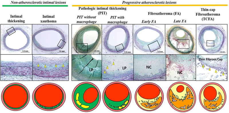
    1. Yahagi K, Kolodgie FD, Otsuka F et al. Pathophysiology of native coronary, vein graft, and in-stent atherosclerosis. Nat Rev Cardiol 2016;13:79–98. - PubMed
    1. Otsuka F, Yasuda S, Noguchi T, Ishibashi-Ueda H. Pathology of coronary atherosclerosis and thrombosis. Cardiovasc Diagn Ther 2016;6:396–408. - PMC - PubMed

Publication types

MeSH terms