Review
. 2020 Oct:75:100895.
doi: 10.1016/j.mam.2020.100895. Epub 2020 Sep 1.

Iron at the host-microbe interface

Affiliations
Review

Iron at the host-microbe interface

Romana R Gerner et al. Mol Aspects Med. 2020 Oct.

Abstract

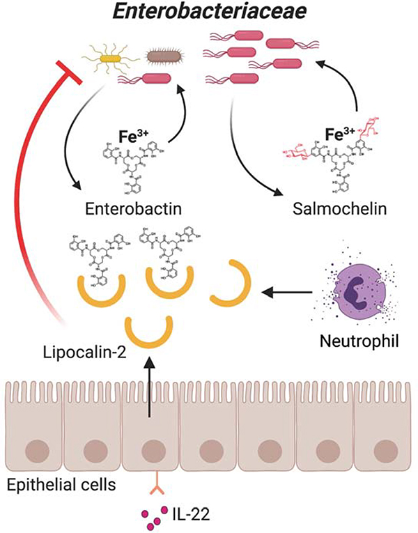
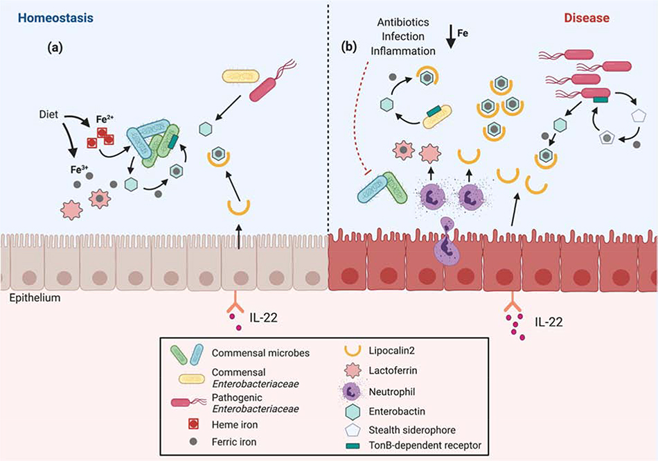
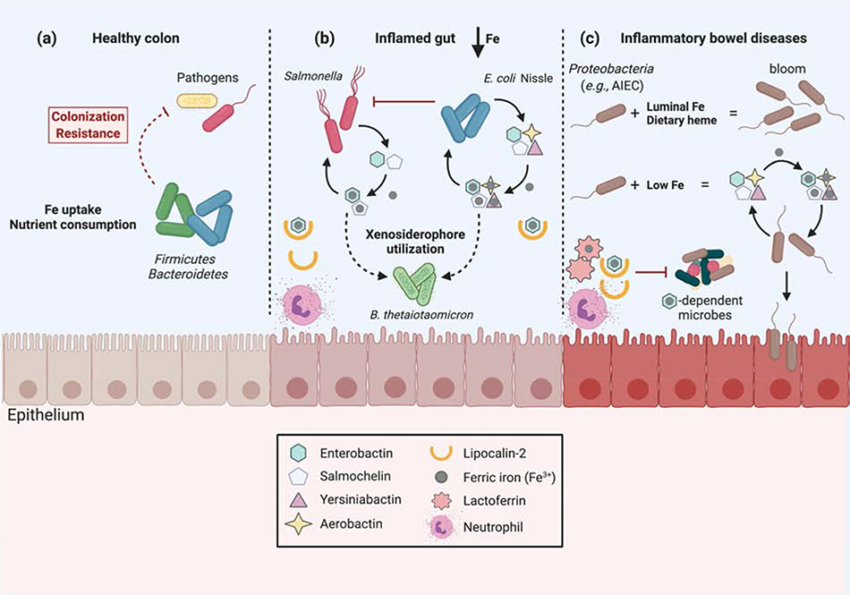
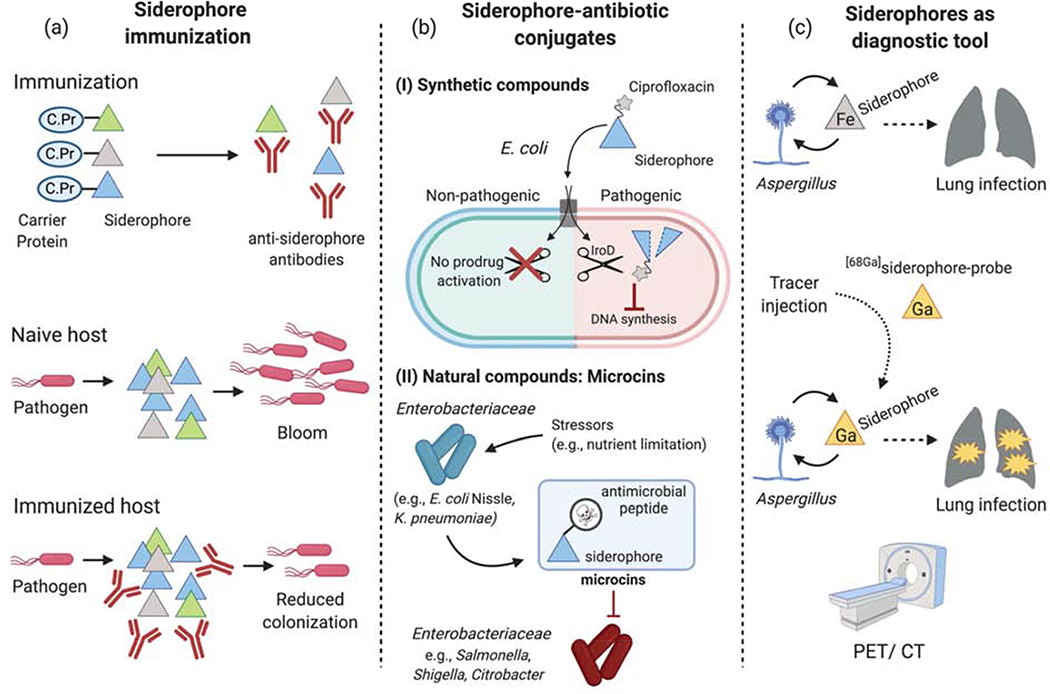
Iron is an essential micronutrient for nearly all living organisms. In addition to facilitating redox reactions, iron is bound by metalloproteins that participate in a variety of biological processes. As the bioavailability of free iron in host environments is extremely low, iron lies at the center of a battle for nutrients between microbes and their host. Mucosal surfaces such as the respiratory and gastrointestinal tracts are constantly exposed to commensal and pathogenic microorganisms. Whereas a key strategy of mammalian antimicrobial defense is to deprive microbes of iron, pathogens and some commensals have evolved effective strategies to circumvent iron limitation. Here we provide an overview of mechanisms underpinning the tug-of-war for iron between microbes and their host, with a particular focus on mucosal surfaces.


Figures

Figure 1.
Figure 1.. Evasion of Lcn2-mediated antimicrobial activity.
Members of the family Enterobacteriaceae can secrete siderophores such as enterobactin and salmochelin into the intestinal lumen to scavenge iron from the host environment. Some immune cells (e.g., neutrophils) and epithelial cells (in response to cytokines including interleukin-22) produce Lcn2, which sequesters enterobactin and thus limits the growth of enterobactin-dependent bacteria by depriving them of iron. In contrast, salmochelin and other ‘stealth’ siderophores enable certain bacteria (mostly pathogens) to evade Lcn2-dependent iron limitation.
Figure 2.
Figure 2.. Model of host-microbe competition for iron in the gut during homeostasis and disease.
(a) Under homeostatic conditions, members of the commensal microbiota acquire iron in the form of heme or by scavenging ferric iron with siderophores such as enterobactin. Low levels of antimicrobial proteins including lactoferrin (Lf) and lipocalin-2 (Lcn2) are present in the gut lumen. Lf sequesters luminal ferric iron, whereas Lcn2 sequesters iron-laden enterobactin. (b) Antibiotic treatment, infection and inflammation can induce IL-22-mediated secretion of Lcn2 from intestinal epithelial cells, and of Lcn2 and Lf from neutrophils that are recruited to the gut. The consequent iron limitation, in turn, induces commensals and pathogens to upregulate various iron uptake mechanisms, including increasing production and secretion of siderophores. Many enteric pathogens secrete stealth siderophores that promote evasion of lipocalin-2-mediated iron sequestration, thus enabling these pathogens to outcompete the gut microbiota and thrive.
Figure 3.
Figure 3.. Iron acquisition by pathogens and commensals.
(a) In the healthy gut, the microbiota is primarily comprised of obligate anaerobes (the phyla Firmicutes and Bacteroidetes). These commensals occupy local niches, consume luminal nutrients and uptake iron (Fe), thereby providing colonization resistance to pathogenic microbes. (b) Intestinal inflammation causes a dramatic reduction in luminal Fe content. The enteric pathogen Salmonella is able to scavenge Fe3+ in this environment, due to the production of salmochelin. However, secretion of multiple siderophores enables the probiotic bacterium E. coli Nissle 1917 to outcompete Salmonella. Moreover, the commensal bacterium Bacteroides thetaiotaomicron sustains colonization during inflammation by utilizing xenosiderophores, a critical mechanism to restore its original colonization state after inflammation is resolved. (c) IBD is characterized by chronic relapsing or remittent intestinal inflammation. During colitis, the increased secretion of host antimicrobial proteins lactoferrin (Lf) and lipocalin-2 (Lcn2) limits the availability of iron to gut microbes. However, the low Fe content induces the upregulation of bacterial Fe uptake genes, which contributes to the blooms of Proteobacteria frequently observed. Even high luminal Fe content (e.g., due to Fe supplementation) promotes the growth of Proteobacteria in the inflamed gut. In patients with Crohn’s disease, adherent-invasive E. coli (AIEC) strains express a variety of Fe-uptake genes, which contribute to the pathogen’s growth during colitis.
Figure 4.
Figure 4.. Siderophores as therapeutic targets and diagnostic tools.
(a) During infection, various siderophores are secreted by pathogens to thrive in the host despite host-mediated iron limitation. In experimental models, siderophores have been targeted for vaccination by linkage to a carrier protein in order to generate anti-siderophore antibodies. Whereas pathogens (e.g., Salmonella, UPEC) were able to successfully replicate in naïve hosts, siderophore-based immunization reduced their colonization levels. (b) Conjugation of antimicrobial peptides or antibiotics to siderophores. (b.I) An enterobactin-ciprofloxacin conjugate is transported through a common siderophore receptor found in the outer membrane of all E. coli strains. Once in the cytoplasm, the IroD esterase, which is mainly found in pathogenic E. coli strains, processes the enterobactin-ciprofloxacin prodrug and thereby enables ciprofloxacin-mediated inhibition of DNA synthesis. (b.II) Certain strains of Enterobacteriaceae, such as E. coli Nissle or K. pneumoniae, secrete microcins in response to nutrient (e.g., Fe) limitation. It is generally thought that the siderophore moiety enables these small antimicrobial peptides to better target susceptible strains of Enterobacteriaceae by utilizing their siderophore receptors for import. (c) Microbial siderophore uptake during infection can be leveraged for imaging applications. During lung infection, Aspergillus scavenges iron (Fe) via siderophores. Injection of siderophores radiolabeled with 68Ga in experimental Aspergillus infections enabled the visualization of the lung infection site by PET/CT.

References

    1. Almeida RS, Wilson D, and Hube B (2009). Candida albicans iron acquisition within the host. FEMS Yeast Res. 9, 1000–1012. - PubMed
    1. Archibald F (1983). Lactobacillus plantarum, an organism not requiring iron. FEMS Microbiol. Lett. 19, 29–32.
    1. Bachman MA, Oyler JE, Burns SH, Caza M, Lépine F, Dozois CM, and Weiser JN (2011). Klebsiella pneumoniae Yersiniabactin Promotes Respiratory Tract Infection through Evasion of Lipocalin 2. Infect. Immun. 79, 3309–3316. - PMC - PubMed
    1. Bachman MA, Lenio S, Schmidt L, Oyler JE, and Weiser JN (2012). Interaction of Lipocalin 2, Transferrin, and Siderophores Determines the Replicative Niche of Klebsiella pneumoniae during Pneumonia. MBio 3. - PMC - PubMed
    1. Bairwa G, Jung WH, and Kronstad JW (2017). Iron acquisition in fungal pathogens of humans. Met. Integr. Biometal Sci. 9, 215–227. - PMC - PubMed

Publication types