Review
. 2020 Oct;41(10):1777-1785.
doi: 10.3174/ajnr.A6739. Epub 2020 Sep 3.

Middle Meningeal Artery: Anatomy and Variations

Affiliations
Review

Middle Meningeal Artery: Anatomy and Variations

S Bonasia et al. AJNR Am J Neuroradiol. 2020 Oct.

Abstract

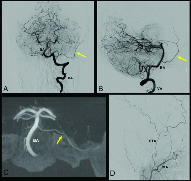
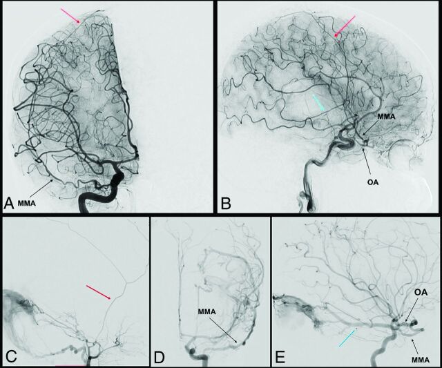
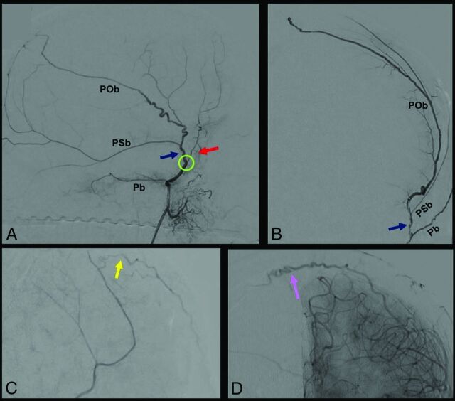
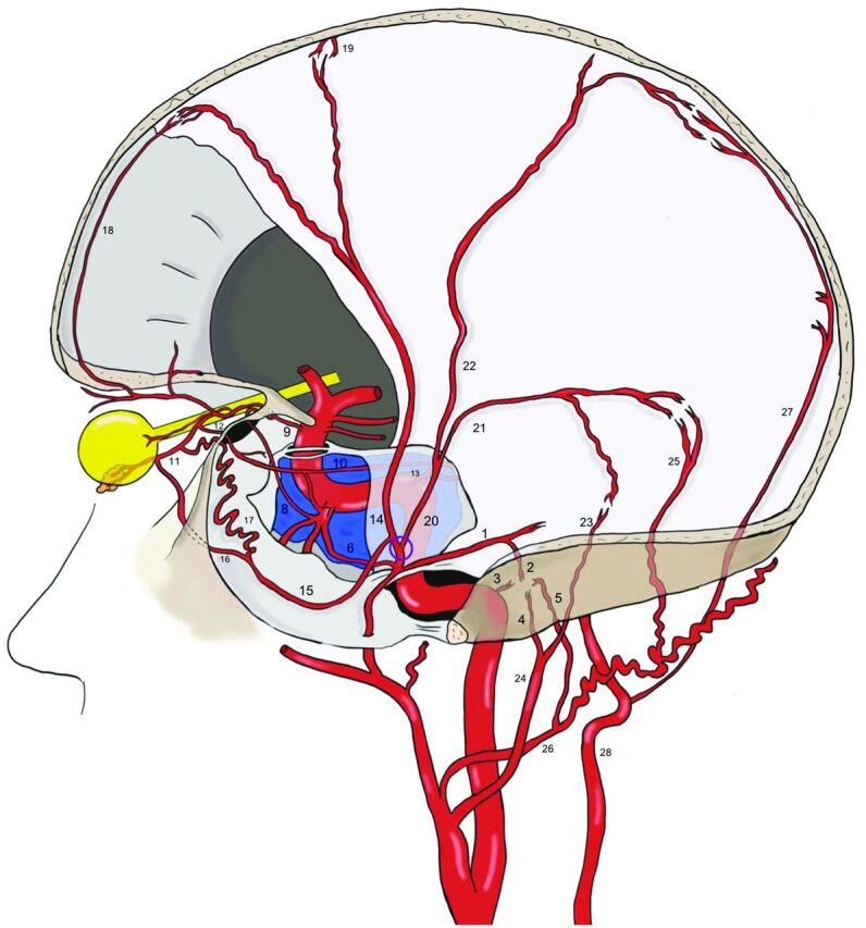
The middle meningeal artery is the major human dural artery. Its origin and course can vary a great deal in relation, not only with the embryologic development of the hyostapedial system, but also because of the relationship of this system with the ICA, ophthalmic artery, trigeminal artery, and inferolateral trunk. After summarizing these systems in the first part our review, our purpose is to describe, in this second part, the anatomy, the possible origins, and courses of the middle meningeal artery. This review is enriched by the correlation of each variant to the related embryologic explanation as well as by some clinical cases shown in the figures. We discuss, in conclusion, some clinical conditions that require detailed knowledge of possible variants of the middle meningeal artery.


Figures

FIG 1.
FIG 1.
A case of MMA origin from basilar artery (BA) pontine perforating branch. In this case, the MMA originates from a pontine branch of the BA, as indicated by the yellow arrow in A, B, and C. A and B, A frontal and lateral view of left vertebral artery (VA) injection, respectively. C, A frontal XperCT (Philips Healthcare) reconstruction with the same MMA origin. D, A distal external carotid artery injection, where the superficial temporal artery (STA) and the IMA are visible, without the typical MMA origin from the IMA.
FIG 2.
FIG 2.
Complete and partial MMA origin from OA. The anteroposterior and lateral view angiograms (A and B respectively) show a rare case of a complete MMA origin from the OA. The OA, through the superficial recurrent OA, gives origin to the MMA, that passes through the lateral part of the superior orbital fissure, and bifurcates into its anterior (red arrow) and posterior division (blue arrow). In the angiograms C, D, and E, a rare case of a partial origin of the MMA from the OA is shown. D and E, Angiograms of a left ICA injection in the frontal and lateral views, where the posterior branch of the MMA (blue arrow) originates at the OA and feeds a tentorial AVF. After the external carotid artery injection (C), only the anterior branch of the MMA is enhanced (red arrow).
FIG 3.
FIG 3.
DSA, showing segments and branches of the MMA. A and B, show selective MMA injection from a lateral (A) and frontal (B) view. The MMA bifurcates at the pterional region (green circle) into 2: an anterior (red arrow) and posterior (blue arrow) division. Before its bifurcation, the MMA gives the petrosal branch (Pb), which courses on the petrous apex. The posterior division gives 2 principal branches: the petrosquamosal branch (PSb) and the parieto-occipital branch (POb). The anterior division ends with 2 kinds of terminal branches, visible after common carotid artery injection: the falcine arteries (yellow arrow) (C), which anastomose with branches of the anterior falcine artery from the OA, and contralateral branches (purple arrow) (D) that cross the midline to anastomose with a contralateral MMA.
FIG 4.
FIG 4.
Anastomoses of the MMA. Before the MMA bifurcation (purple circle), the petrous branch (1), from which the superior tympanic artery (2) originates, anastomoses into the middle ear with the caroticotympanic artery (3, from the ICA), and with the inferior tympanic artery (4, from the ascending pharyngeal artery), with the posterior tympanic artery (5, from the occipital artery). The cavernous branch of the MMA (6) on the other side anastomoses with the inferolateral trunk (ILT) (7), which is itself connected to the OA (9) through the deep recurrent OA (8). The ILT, the MMA, and the OA are also linked to each other through the marginal tentorial artery (10), whose origin can vary from the lacrimal artery (11), via superficial recurrent OA (12), to the meningohypophyseal trunk (13). After the MMA bifurcation at the pterion, its frontal division (14) gives a medial branch (15), which can bifurcate intracranially into a lateral meningolacrimal artery (16), and a medial sphenoidal artery (17). Both branches reach the lacrimal artery, even if the meningolacrimal artery more distal than the sphenoidal artery. The anastomoses with the OA and the ILT represent the most dangerous connections in the case of MMA transarterial embolization because of the risk of particle embolism into these arteries. The frontal division of the MMA reaches the convexity, following the coronal suture and anastomoses with the anterior falcine artery (18, OA–anterior ethmoidal artery) and with branches of the contralateral MMA (19). The posterior division of the MMA (20) divides into a petrosquamosal branch (21) and a parieto-occipital branch (22). The former anastomoses with the jugular branch (23) of the ascending pharyngeal artery (24) and with the mastoid branch (25) of the occipital artery (26). The latter is linked to the posterior meningeal artery (27), from the vertebral artery (28) at the border areas.

References

    1. Martins C, Yasuda A, Campero A, et al. . Microsurgical anatomy of the dural arteries. Neurosurgery 2005;56:211–51; discussion 211–51 10.1227/01.neu.0000144823.94402.3d - DOI - PubMed
    1. Merland JJ, Theron J, Lasjaunias P, et al. . Meningeal blood supply of the convexity. J Neuroradiol 1977;4:129–74 - PubMed
    1. Lasjaunias P, Bereinstein A, ter Brugge K. Surgical Neuroangiography KG. Berlin: Springer-Verlag; 2001
    1. Low FN. An anomalous middle meningeal artery. Anat Rec 1946;95:347–51 10.1002/ar.1090950310 - DOI - PubMed
    1. Altmann F. Anomalies of the internal carotid artery and its branches; their embryological and comparative anatomical significance; report of a new case of persistent stapedial artery in man. Laryngoscope 1947;57:313–39 10.1288/00005537-194705000-00002 - DOI - PubMed

MeSH terms

LinkOut - more resources