. 2020 Oct;41(10):1841-1848.
doi: 10.3174/ajnr.A6758. Epub 2020 Sep 3.

Transcranial MR Imaging-Guided Focused Ultrasound Interventions Using Deep Learning Synthesized CT

Affiliations

Transcranial MR Imaging-Guided Focused Ultrasound Interventions Using Deep Learning Synthesized CT

P Su et al. AJNR Am J Neuroradiol. 2020 Oct.

Abstract

Background and purpose: Transcranial MR imaging-guided focused ultrasound is a promising novel technique to treat multiple disorders and diseases. Planning for transcranial MR imaging-guided focused ultrasound requires both a CT scan for skull density estimation and treatment-planning simulation and an MR imaging for target identification. It is desirable to simplify the clinical workflow of transcranial MR imaging-guided focused ultrasound treatment planning. The purpose of this study was to examine the feasibility of deep learning techniques to convert MR imaging ultrashort TE images directly to synthetic CT of the skull images for use in transcranial MR imaging-guided focused ultrasound treatment planning.

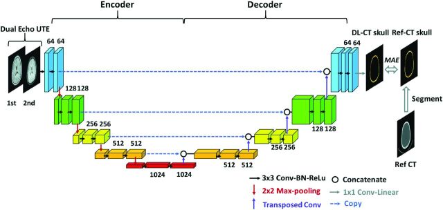
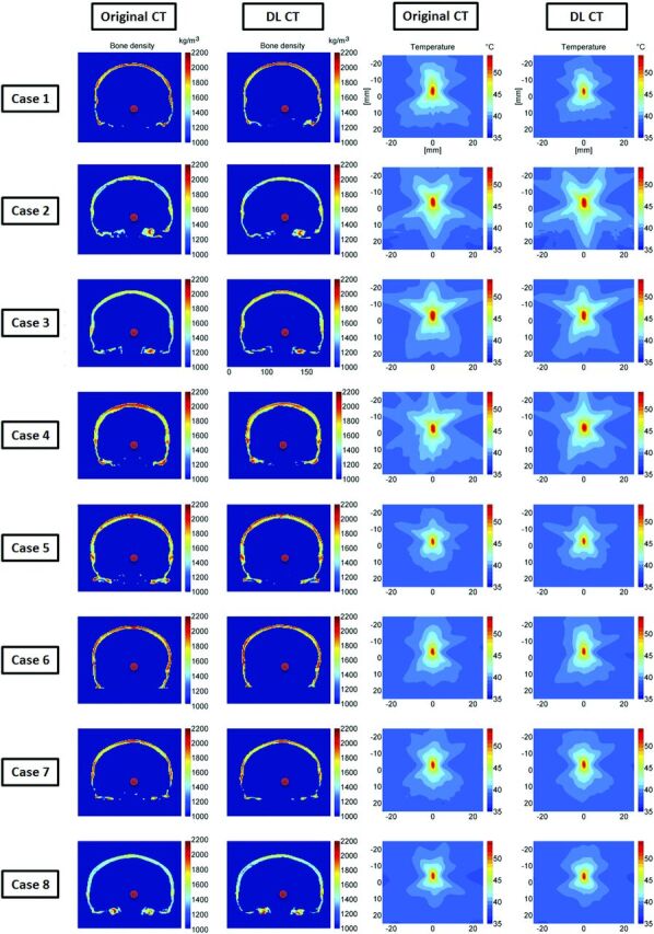
Materials and methods: The U-Net neural network was trained and tested on data obtained from 41 subjects (mean age, 66.4 ± 11.0 years; 15 women). The derived neural network model was evaluated using a k-fold cross-validation method. Derived acoustic properties were verified by comparing the whole skull-density ratio from deep learning synthesized CT of the skull with the reference CT of the skull. In addition, acoustic and temperature simulations were performed using the deep learning CT to predict the target temperature rise during transcranial MR imaging-guided focused ultrasound.

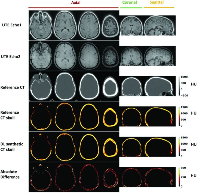
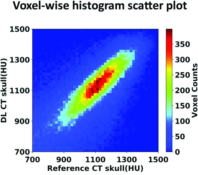
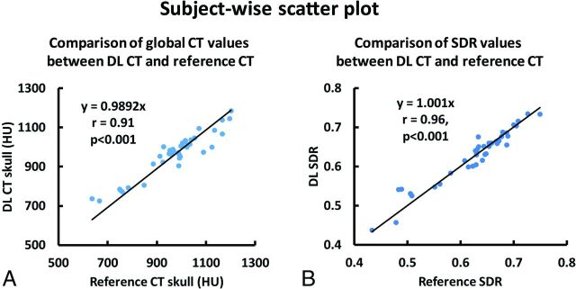
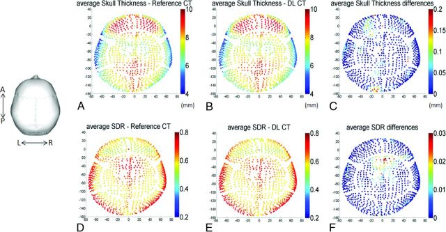
Results: The derived deep learning model generates synthetic CT of the skull images that are highly comparable with the true CT of the skull images. Their intensities in Hounsfield units have a spatial correlation coefficient of 0.80 ± 0.08, a mean absolute error of 104.57 ± 21.33 HU, and a subject-wise correlation coefficient of 0.91. Furthermore, deep learning CT of the skull is reliable in the skull-density ratio estimation (r = 0.96). A simulation study showed that both the peak target temperatures and temperature distribution from deep learning CT are comparable with those of the reference CT.

Conclusions: The deep learning method can be used to simplify workflow associated with transcranial MR imaging-guided focused ultrasound.


Figures

FIG 1.
FIG 1.
Schema of the employed deep learning architecture based on the widely used U-Net convolutional neural network consisting of encoding and decoding pathways. Dual-echo UTE images were used as the input for the network. Reference CT of the skull was segmented from the reference CT and was used as the predication target. The difference between output of the network, DL synthetic CT of the skull, and reference CT of the skull was minimized using MAE loss function. Drop-out regularization (rate = 0.5) was applied connecting the encoder and decoder. BN indicates batch normalization; ReLu, rectified linear unit; Conv, convolutional layer; Ref-CT, reference CT.
FIG 2.
FIG 2.
Deep learning results from 1 representative testing subject (55 years of age, female). From top to bottom: UTE echo 1 and echo 2 images, reference CT, segmented reference CT of the skull, DL synthetic CT of the skull, and the absolute difference between the 2. For this subject, the Dice coefficient for skull masks between DL synthetic CT and the reference CT is 0.92, and the mean absolute difference is 96.27 HU.
FIG 3.
FIG 3.
Voxelwise 2D histogram scatterplot between the reference skull CT intensity and the DL synthetic skull CT signal intensity in Hounsfield units within skull. The correlation coefficient is r = 0.80 (same testing subject as in Fig 2). Color bar represents the voxel count.
FIG 4.
FIG 4.
A, Association of average CT Hounsfield unit values between the DL synthetic CT of the skull and the reference CT of the skull for all 40 testing subjects from cross-validation. Each dot represents 1 subject. B, The relationship between SDR values determined from the reference CT and DL synthetic CT from all 40 subjects. Each dot represents 1 subject.
FIG 5.
FIG 5.
A and B, The calculated average skull thickness map from the reference CT and the DL synthetic CT images, respectively, from all 40 testing subjects. C, The differences between A and B with a maximum thickness difference of 0.2 mm (2% error) and the average error of 0.03 mm (0.3%). Note that regional maps are based on the entries of the 1024 ultrasound beams from the ExAblate system (InSightec). D and E, The calculated average SDR map based on the reference CT and DL synthetic CT images from all 40 subjects. F, The differences between D and E with a maximum SDR difference of 0.03 (4% error) and the average error of 1024 entries was <0.01 (1.3%).
FIG 6.
FIG 6.
Comparison of calculated bone density and the simulated temperature rise. The first and second columns show the calculated bone density map using the reference CT and DL synthetic CT images on 8 representative testing cases, in which the red dots are the assigned focal targets. In the third and fourth columns, the simulated temperature elevations at the focal spots caused by a 16-second, 1000-W sonication are compared between reference CT and DL synthetic CT on a base brain temperature of 37°C. The simulated peak temperature rise values based on original and DL synthetic CT images for all 8 subjects are the following: case 1: 55.3°C and 54.2°C (6.0% error on temperature rise), case 2: 59.0°C and 58.9°C (0.5% error), case 3: 57.6°C and 55.9°C (8.2% error), case 4: 57.5°C and 59.1°C (7.8% error), case 5: 54.3°C and 54.6°C (0.6% error), case 6: 55.5°C and 56.0°C (0.9% error), case 7: 54.5°C and 55.1°C (1.1% error), case 8: 53.5°C and 54.5°C (1.9% error), respectively. These errors are well within the errors that one might expect from the simulation.

Similar articles

Cited by

References

    1. Elias WJ, Huss D, Voss T, et al. . A pilot study of focused ultrasound thalamotomy for essential tremor. N Engl J Med 2013;369:640–48 10.1056/NEJMoa1300962 - DOI - PubMed
    1. Jeanmonod D, Werner B, Morel A, et al. . Transcranial magnetic resonance imaging-guided focused ultrasound: noninvasive central lateral thalamotomy for chronic neuropathic pain. Neurosurg Focus 2012;32:E1 10.3171/2011.10.FOCUS11248 - DOI - PubMed
    1. Monteith S, Sheehan J, Medel R, et al. . Potential intracranial applications of magnetic resonance-guided focused ultrasound surgery. J Neurosurg 2013;118:215–21 10.3171/2012.10.JNS12449 - DOI - PubMed
    1. Guo S, Zhuo J, Li G, et al. . Feasibility of ultrashort echo time images using full-wave acoustic and thermal modeling for transcranial MRI-guided focused ultrasound (tcMRgFUS) planning. Phys Med Biol 2019;64:095008 10.1088/1361-6560/ab12f7 - DOI - PubMed
    1. Wiesinger F, Sacolick LI, Menini A, et al. . Zero TE MR bone imaging in the head. Magn Reson Med 2016;75:107–14 10.1002/mrm.25545 - DOI - PubMed