. 2020 Dec;19(12):1986-1997.
doi: 10.1074/mcp.RA120.002212. Epub 2020 Sep 3.

A Comprehensive Gender-related Secretome of Plasmodium berghei Sexual Stages

Affiliations

A Comprehensive Gender-related Secretome of Plasmodium berghei Sexual Stages

Felicia Grasso et al. Mol Cell Proteomics. 2020 Dec.

Abstract

Plasmodium, the malaria parasite, undergoes a complex life cycle alternating between a vertebrate host and a mosquito vector of the genus Anopheles In red blood cells of the vertebrate host, Plasmodium multiplies asexually or differentiates into gamete precursors, the male and female gametocytes, responsible for parasite transmission. Sexual stage maturation occurs in the midgut of the mosquito vector, where male and female gametes egress from the host erythrocytes to fuse and form a zygote. Gamete egress entails the successive rupture of two membranes surrounding the parasite, the parasitophorous vacuole membrane and the erythrocyte plasma membrane. In this study, we used the rodent model parasite Plasmodium berghei to design a label-free quantitative proteomic approach aimed at identifying gender-related proteins differentially released/secreted by purified mature gametocytes when activated to form gametes. We compared the abundance of molecules secreted by wild type gametocytes of both genders with that of a transgenic line defective in male gamete maturation and egress. This enabled us to provide a comprehensive data set of egress-related molecules and their gender specificity. Using specific antibodies, we validated eleven candidate molecules, predicted as either gender-specific or common to both male and female gametocytes. All of them localize to punctuate, vesicle-like structures that relocate to cell periphery upon activation, but only three of them localize to the gametocyte-specific secretory vesicles named osmiophilic bodies. Our results confirm that the egress process involves a tightly coordinated secretory apparatus that includes different types of vesicles and may put the basis for functional studies aimed at designing novel transmission-blocking molecules.

Keywords: Parasite; Plasmodium berghei; blood; egress; gametocyte; infectious disease; label-free quantification; malaria; mass spectrometry; osmiophilic bodies; pathogens; secretome.


Conflict of interest statement

Conflict of interest—Authors declare no competing interests.

Figures
None
Graphical abstract
Fig. 1.
Fig. 1.
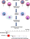
Design of proteomic experiments. P. berghei mature gametocytes were isolated from peripheral blood of mice infected with a wt parasite line or a transgenic line lacking the actin II gene, actII(-), defective in male gametogenesis and egress. Gametocytes were activated in vitro by a drop in temperature and the addition of xanthurenic acid (XA). Proteins released by either male or female gametocytes were identified by comparative label-free quantitative proteomics.
Fig. 2.
Fig. 2.
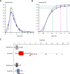
Abundance ratio distributions of wt/actII(-) and wt/g377(-). A, Top3 ratios of wt (light blue), actII(-) (pink) and g377(-) (green) follow the same log-normal distribution according with the Kolmogorov-Smirnov test (p > 0.05). The common median equal to 1 is indicated as a dotted line. B, Cumulative distribution of Top3 ratio values. Dotted lines indicate the percentage of values falling within one standard deviation from the median (95.8% for wt; 91.2% for actII(-) and 85.3% for g377(-)). C, Box plot representation of Top3 ratio value distributions of wt/actII(-) and wt/g377(-) and reference sets A (wt and actII(-) Top3 ratios) and B (wt and g377(-) Top3 ratios). Median value of wt/actI(-) is twice that of reference set A, being male proteome under-represented in actII(-) egress secretome. Red arrowhead indicates the maximum value of 6.1, at the boundary of the highest quartile of wt/actII(-) distribution, whereas red dots represent abundance ratio values external to the whisker identifying male-related candidates. Abundance ratio values of these selected candidates fall within the distribution range when wt/g377(-) or the reference B are considered.
Fig. 3.
Fig. 3.
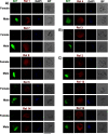
Gender-specific expression of candidate proteins selected from gametocyte secretome. Expression profile of selected molecules was analyzed by specific antibodies in double IFA on fixed wt gametocytes. Gender specificity was determined using an immune serum against the nuclear protein SET, highly abundant in male gametocytes. A, proteins specifically expressed in male gametocytes; B, proteins specifically expressed in female gametocytes; C, proteins expressed in both genders. Nuclei are stained with DAPI. BF, bright field. Scale bar indicates 5 μm.
Fig. 4.
Fig. 4.
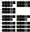
Subcellular localization of candidate proteins. Subcellular localization of the selected proteins was evaluated by double IFA with immune sera raised against the OB markers G377, expressed only in female gametocytes, and MDV1 expressed in both genders. A, Rel 1, Rel 14 and Rel 16 immune sera stain OBs. B, All the other immune sera tested stain vesicle-like structures distinct from OBs. Nuclei are stained with DAPI. BF, bright field. Scale bar indicates 5 μm.
Fig. 5.
Fig. 5.
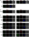
Immune localization of MOB-associated proteins. Double IFA with antibodies against the MOB-resident MiGS and PbSUB1 shows that the two proteins localize to the same vesicles both in noninduced and induced male gametocytes. Nuclei are stained with DAPI. Scale bar indicates 5 μm.
Fig. 6.
Fig. 6.
Subcellular localization of candidate proteins in activated gametocytes. Rel antibodies against male- (A) and female (B)-specific proteins or against proteins detected in both genders (C) were used in IFA on fixed gametocytes activated for 5–8 min. Female gametocytes were specifically stained by anti-G377 antibodies, whereas male gametocytes by anti-Tubulin antibodies. Nuclei are stained with DAPI. BF, bright field. Scale bar indicates 5 μm.

References

    1. Bousema T., and Drakeley C. (2011) Epidemiology and infectivity of Plasmodium falciparum and Plasmodium vivax gametocytes in relation to malaria control and elimination. Clin. Microbiol. Rev. 24, 377–410 - PMC - PubMed
    1. Schneider P., Bousema J. T., Gouagna L. C., Otieno S., van de Vegte-Bolmer M., Omar S. A., and Sauerwein R. W. (2007) Submicroscopic Plasmodium falciparum gametocyte densities frequently result in mosquito infection. Am. J. Trop. Med. Hyg. 76, 470–474 - PubMed
    1. Garcia G. E., Wirtz R. A., Barr J. R., Woolfitt A., and Rosenberg R. (1998) Xanthurenic acid induces gametogenesis in Plasmodium, the malaria parasite. J. Biol. Chem. 273, 12003–12005 - PubMed
    1. Kuehn A., and Pradel G. (2010) The coming-out of malaria gametocytes. J. Biomed. Biotechnol. 2010, 976827. - PMC - PubMed
    1. Sologub L., Kuehn A., Kern S., Przyborski J., Schillig R., and Pradel G. (2011) Malaria proteases mediate inside-out egress of gametocytes from red blood cells following parasite transmission to the mosquito. Cell Microbiol. 13, 897–912 - PubMed

Publication types

LinkOut - more resources