. 2020 Sep 4;369(6508):1220-1227.
doi: 10.1126/science.aba4904.

Structure of a human 48 S translational initiation complex

Affiliations

Structure of a human 48 S translational initiation complex

Jailson Brito Querido et al. Science. .

Abstract

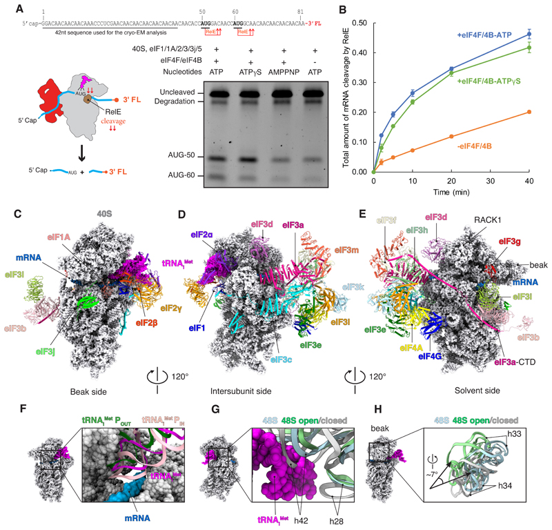
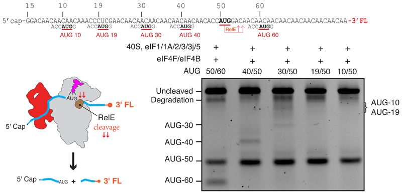
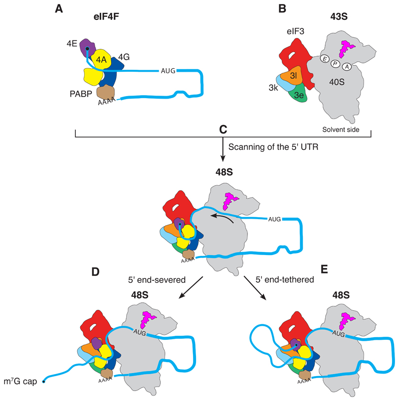
A key step in translational initiation is the recruitment of the 43S preinitiation complex by the cap-binding complex [eukaryotic initiation factor 4F (eIF4F)] at the 5' end of messenger RNA (mRNA) to form the 48S initiation complex (i.e., the 48S). The 48S then scans along the mRNA to locate a start codon. To understand the mechanisms involved, we used cryo-electron microscopy to determine the structure of a reconstituted human 48S The structure reveals insights into early events of translation initiation complex assembly, as well as how eIF4F interacts with subunits of eIF3 near the mRNA exit channel in the 43S The location of eIF4F is consistent with a slotting model of mRNA recruitment and suggests that downstream mRNA is unwound at least in part by being "pulled" through the 40S subunit during scanning.


Conflict of interest statement

Competing interests: The authors declare no competing interests.

Figures

Fig. 1
Fig. 1. Structure and functional characterization of human 48S complex.
(A) RelE cleavage shows that the efficiency of start-site selection depends on eIF4F (lanes 1 and 4) as well as on ATP hydrolysis (lines 1 and 3). The slowly hydrolysable ATP analog ATPγS (lane 2) has almost the same efficiency of start-site selection when compared with ATP. For the gel, RelE cleavage assay was performed 20 min after the formation of the 48S complex. The degradation band is present in the mRNA prep even in the absence of RelE cleavage (fig. S1A). FL, fluorescent labeled; nt, nucleotide. (B) Kinetic analysis in the presence of eIF4F with ATP or ATPgS, and with ATP absence of eIF4F, shows an eIF4F and ATP-dependent mechanism of scanning under our experimental conditions, and that ATPγS is almost as good as ATP. Error bars indicate SEM. (C to E) Overall structure of 48S shown in different orientations. eIFs are shown in cartoon. tRNAiMet and 40S are represented as magenta and gray spheres. Sugar phosphate backbone of the mRNA is shown as a blue surface. CTD, C-terminal domain. (F) Superposition of the tRNAi Met with the structure of tRNAi Met in the context of 48S in open (POUT) and closed conformation (PIN) (14) shows an intermediate conformation of the anticodon stem loop. (G and H) Superposition of 18S rRNA with the structure of 48S in the open and closed conformations (14) to highlight the changes in their conformation and the swivel movement of the head during scanning.
Fig. 2
Fig. 2. Interactions of eIF3 with mRNA and 40S.
(A) Close-up of the mRNA exit site highlighting the interaction of eIF3a (pink) with mRNA (blue sugar-phosphate backbone). K, Lys. (B) eIF3a interacts with ribosomal protein eS1 (yellow) near the exit site. Red dashed lines represent protein-protein or protein-RNA interactions. eIF3a R14-a and K23 interact with eS1 E78 and S192. Furthermore, eIF3a K63 interacts with rRNA ES7S C1116. C, Cys; E, Glu; K, Lys; R, Arg; S, Ser. (C) Close-up of R14 in eIF3a to highlight in two alternative rotamer conformations (R14-a and R14-b) and the interaction of R14-b with D77 of r-protein eS1. D, Asp; Q, Gln. (D) Close-up of the interaction of eIF3c with 18S rRNA on the back of the 40S. G, Gly. (E) eIF3d-NTT (orchid) fitted into the cryo-EM map to highlight the close interactions with PCI domains of eIF3c (cyan surface) and eIF3e (green surface). V, Val. (F) Close-up of the PCI domain of eIF3c to highlight some described interactions with eIF3d-NTT. A, Ala; W, Trp.
Fig. 3
Fig. 3. Interactions of peripheral subunits of eIF3.
(A) eIF3g-RNA binding motif viewed from solvent side to highlight its interaction with rRNA in helix 16 and ribosomal proteins uS3 and eS10. (B) Binding interface between eIF3g and the ribosomal protein uS3 at the mRNA entry channel. H, His; I, Ile; L, Leu; Y, Tyr. (C) eIF3j binds to ribosomal proteins eS30 and uS12 near the A site. The C-terminal tail of eIF3j (eIF3j-CTT) interacts with ribosomal rRNA in helix 34. eIF3j-NTT is positioned next to the GTPase binding region of the 40S. (D) Superposition of eIF3j with the structure of ABCE1 (PDB: 5LL6) bound to the 40S subunit (post splitting) (29). (E and F) eIF3j-NTT extends toward the GTPase binding region of the 40S, where the nucleotide binding domain 1 (NBD1) of ABCE1 binds.
Fig. 4
Fig. 4. Interactions between eIF4F and eIF3 octameric structural core.
(A and B) Rigid-body fitting (correlation = 0.92) of human homology model of eIF4A/eIF4G-HEAT1 into a cryo-EM map filtered to local resolution (6 to 11 Å). (C) eIF4A binds to a pocket formed by eIF3l and eIF3e. (D) Saturation binding curves showing the fraction of eIF4A bound to eIF3 in the presence or absence of eIF4G. Error bars indicate SEM. (E) Surface representation of the mRNA (sugar-phosphate backbone) to highlight the path in the 48S. Blue dots represent a tentative path for the mRNA from the exit site (position −14 from the P site) toward eIF4A-NTD. The tentative path is based on weak unassigned density (fig. S16).
Fig. 5
Fig. 5. Blind spot for start-site recognition.
RelE cleavage assay (materials and methods) shows a blind spot for start-site selection for mRNAs with a start codon less than 30 to 40 nucleotides from the 5′ end. All five mRNAs tested have an AUG at position 50 just beyond the blind spot and a second AUG at positions downstream (60 nucleotides) or upstream (40, 30, 19, and 10 nucleotides) of it. The main site of cleavage is at AUG 50 in all cases, showing that initiation occurs primarily at the first start codon downstream of the blind spot, and there is little or no cleavage at start codons for 10 to 40 nucleotides.
Fig. 6
Fig. 6. Model for mRNA scanning during canonical translational initiation suggested by the structure.
The eIF4F at the m7G cap at the 5′ end of mRNA (A) recruits the 43S complex of the 40S subunit with initiation factors and initiator tRNAi Met (B) to form the 48S complex (C). eIF4F binds to the eIF3 structural core, which places eIF4E 30 to 40 nucleotides upstream of the P site of the 40S ribosomal subunit. During scanning, the mRNA is pulled through the 40S subunit [indicated by arrow in (C)], until the start codon is reached (D and E). In two alternative scenarios, eIF4F could dissociate from the cap during scanning (D), or it could stay bound resulting in the mRNA forming a loop (E). Although not part of the structure in this work, the mRNA is shown with a poly(A) tail and a PABP interacting with eIF4F to reflect the situation in vivo (39).

References

    1. Grifo JA, Tahara SM, Morgan MA, Shatkin AJ, Merrick WC. New initiation factor activity required for globin mRNA translation. J Biol Chem. 1983;258:5804–5810. Medline. - PubMed
    1. Morino S, Imataka H, Svitkin YV, Pestova TV, Sonenberg N. Eukaryotic translation initiation factor 4E (eIF4E) binding site and the middle one-third of eIF4GI constitute the core domain for cap-dependent translation, and the C-terminal one-third functions as a modulatory region. Mol Cell Biol. 2000;20:468–477. doi: 10.1128/MCB.20.2.468-477.2000. Medline. - DOI - PMC - PubMed
    1. Marintchev A, Edmonds KA, Marintcheva B, Hendrickson E, Oberer M, Suzuki C, Herdy B, Sonenberg N, Wagner G. Topology and regulation of the human eIF4A/4G/4H helicase complex in translation initiation. Cell. 2009;136:447–460. doi: 10.1016/j.cell.2009.01.014. Medline. - DOI - PMC - PubMed
    1. Villa N, Do A, Hershey JWB, Fraser CS. Human eukaryotic initiation factor 4G (eIF4G) protein binds to eIF3c, -d, and -e to promote mRNA recruitment to the ribosome. J Biol Chem. 2013;288:32932–32940. doi: 10.1074/jbc.M113.517011. Medline. - DOI - PMC - PubMed
    1. LeFebvre AK, Korneeva NL, Trutschl M, Cvek U, Duzan RD, Bradley CA, Hershey JWB, Rhoads RE. Translation initiation factor eIF4G-1 binds to eIF3 through the eIF3e subunit. J Biol Chem. 2006;281:22917–22932. doi: 10.1074/jbc.M605418200. Medline. - DOI - PMC - PubMed

Publication types

MeSH terms