. 2020 Sep 4;369(6508):1227-1233.
doi: 10.1126/science.abb8330.

A defined structural unit enables de novo design of small-molecule-binding proteins

Affiliations

A defined structural unit enables de novo design of small-molecule-binding proteins

Nicholas F Polizzi et al. Science. .

Abstract

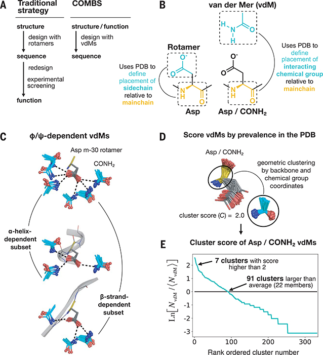
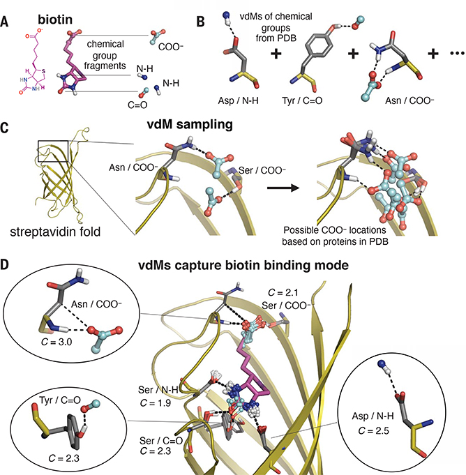
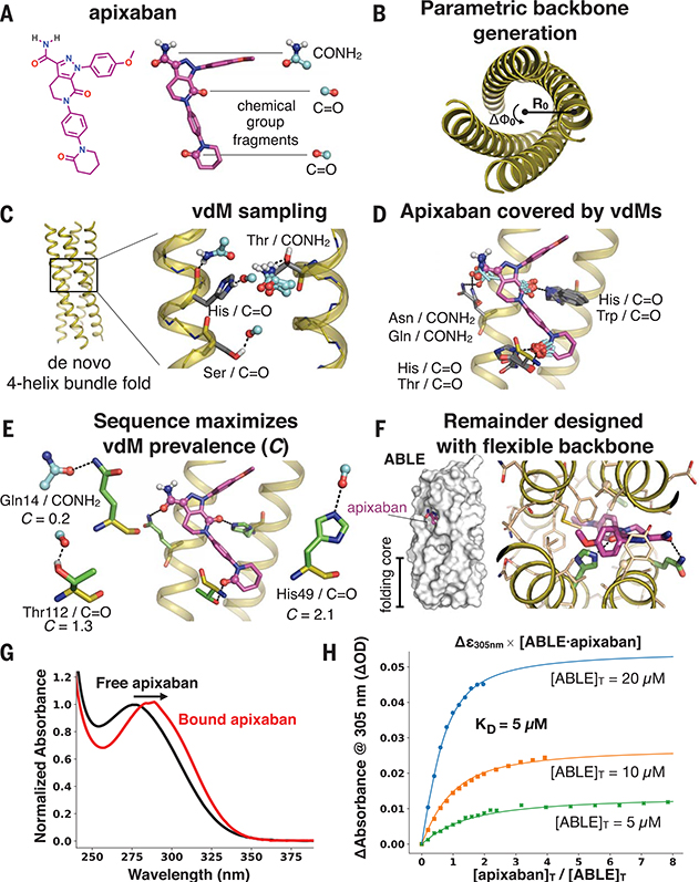
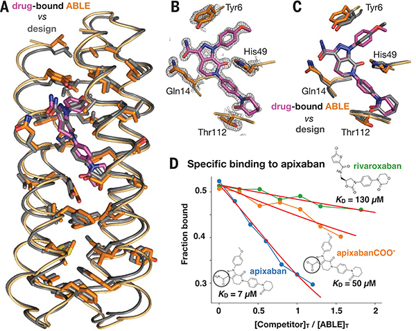
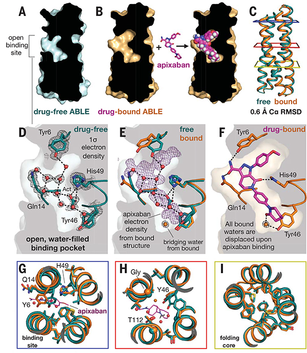
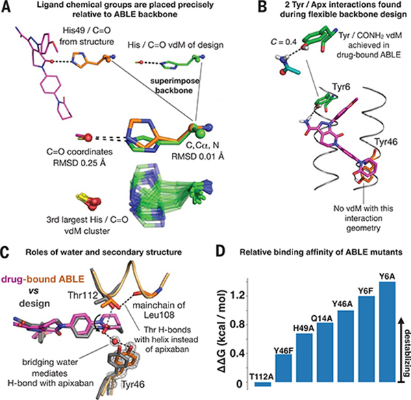
The de novo design of proteins that bind highly functionalized small molecules represents a great challenge. To enable computational design of binders, we developed a unit of protein structure-a van der Mer (vdM)-that maps the backbone of each amino acid to statistically preferred positions of interacting chemical groups. Using vdMs, we designed six de novo proteins to bind the drug apixaban; two bound with low and submicromolar affinity. X-ray crystallography and mutagenesis confirmed a structure with a precisely designed cavity that forms favorable interactions in the drug-protein complex. vdMs may enable design of functional proteins for applications in sensing, medicine, and catalysis.


Figures

Fig. 1.
Fig. 1.. A vdM is a structural unit relating chemical group position to the protein backbone.
(A) Workflow of a traditional protein design strategy versus that of COMBS. (B) Definition of a vdM. A chemical group is interacting if it is in van der Waals contact with the protein side chain or main chain. Like rotamers, vdMs are derived from a large set of high-quality protein crystal structures. A vdM of aspartic acid (Asp) and carboxamide (CONH2, cyan) is shown. (C) vdMs are φ, ψ, and rotamer dependent; this is illustrated by the top vdMs of the m-30 rotamer of Asp, clustered by location of CONH2 after exact superposition of main chain N, Cα, and C atoms. (D and E) We ranked vdMs by prevalence in the PDB, quantified by a cluster score C [the natural logarithm of the ratio of the number of members in a cluster (NvdM) to the average number of members in a cluster (⟨hNvdM⟩)]. The seventh-largest cluster of Asp/CONH2 vdMs is shown as an example in (D).
Fig. 2.
Fig. 2.. Prevalent vdMs describe the binding site of biotin in streptavidin.
(A and B) We constructed vdMs of the polar chemical groups of biotin by searching the PDB for protein interactions with the (i) backbone amide nitrogen (N-H), (ii) backbone carbonyl or carbonyl from Asn or Gln side chains (C=O), and (iii) carboxylate of Asp or Glu side chains (COO). (C) Using the native sequence of streptavidin, vdMs were sampled on the streptavidin backbone to generate possible locations for productive interactions with the chemical groups. Here, Asn and Ser vdMs of COO are sampled at two positions of the backbone. (D) vdMs with chemical groups (cyan) that are nearest neighbors (0.6 Å RMSD) to those of biotin in its binding site are overlaid on top of biotin (purple).
Fig. 3.
Fig. 3.. Apixaban-binding helical bundle (ABLE) design strategy.
(A to F) Steps of the design process. (A) We targeted simultaneous engagement of two carbonyls (C=O) and the carboxamide (CONH2) of apixaban. (B) We computationally generated a set of 32 designable four-helix bundle folds based on a mathematical parameterization. (C) vdM sampling of CONH2 and C=O allowed us to enumerate statistically preferred locations of these chemical groups relative to the backbone. (D) We used a precomputed set of vdMs with apixaban superimposed by one of its chemical groups to position apixaban within the bundle, such that it was guaranteed to have at least one vdM that accommodates its position. Chemical groups of vdMs that overlap with those of apixaban are found by a nearest-neighbors lookup. Multiple vdMs contributing from one residue position are possible, e.g., His/C=O and Trp/C=O vdMs, and can be used in separate designs. (E) Specific choices of vdMs for each chemical group of the ligand were made by maximizing the use of highly enriched vdMs in the binding site (high C score) (Fig. 1, D and E). Final ligand positions and interactions for the six experimentally characterized designs were chosen by maximizing both C and the burial of the apolar surface area of apixaban. The vdMs chosen to comprise the binding site of ABLE are shown along with their cluster scores. (F) The location of apixaban and its vdM-derived interactions with the protein are constrained in a subsequent flexible backbone sequence design protocol. (G) The electronic absorbance spectrum of apixaban is red-shifted upon binding to ABLE. The black spectrum shows apixaban (4 μM) in buffer containing 50 mM NaPi, 100 mM NaCl (pH 7.4). The red spectrum is the difference of the absorbance spectrum of ABLE alone (20 μM) and the spectrum of ABLE (20 μM) with apixaban (4 μM). The spectra were normalized to the peak maximum for comparison. These experiments were facilitated by the high extinction coefficient of apixaban and the lack of Trp in ABLE. (H) Global fit of a single-site binding model to the absorbance changes at 305 nm upon titration of apixaban into 5, 10, and 20 μM solutions of ABLE. The KD from the fit is 5 (± 1) μM, which was confirmed by fluorescence polarization competition experiments (supplementary materials).
Fig. 4.
Fig. 4.. The structure of apixaban-bound ABLE agrees with the design.
(A) Superposition of backbone Cα atoms of structure (protein in orange, apixaban in purple) and design (gray; 0.7 Å RMSD), showing side chains of amino acids in the protein core. (B) ABLE’s binding site from the structure (1.3-Å resolution), showing vdM-derived interactions with apixaban (purple). The 2mFo-DFc composite omit map is contoured at 1.5 σ. The map was generated from a model that omitted coordinates of apixaban. The protein backbone of these residues is shown in cartoon format. (C) Overlay of designed interactions (gray), after the designed model was superimposed onto the Cα atoms of the structure (protein in orange, apixaban in purple). (D) Fluorescence anisotropy competition experiments (485-nm excitation, 528-nm emission) showed that ABLE binds apixaban specifically. The bound fluorophore apixaban–polyethylene glycol–fluorescein isothiocyanate (apixaban-PEG-FITC) (supplementary text and fig. S9) is dislodged by addition of competing ligand. Anisotropy was converted to the fraction bound by use of a one-site binding model (supplementary text). The ABLE concentration was 20 μM, and the apixaban-PEG-FITC concentration was 25 nM in buffer containing 50 mM NaPi, 100 mM NaCl (pH 7.4). Apixaban COO is identical to apixaban except that it contains a carboxylate instead of a carboxamide (circled). Rivaroxaban is another inhibitor that also binds tightly to factor Xa by using the same binding mode as apixaban but shows only very weak binding to ABLE. Fits to a competitive binding model are shown in red. KD values: rivaroxaban, 130 (± 10) μM; apixaban COO, 50 (± 5) μM; apixaban, 7 (± 2) μM.
Fig. 5.
Fig. 5.. Drug-free ABLE has a preorganized structure with an open binding site competent for binding.
(A) A slice through a surface representation of the 1.3-Å resolution structure of unliganded ABLE shows an open binding cavity. (B) Same slice, shown for the structure of apixaban-bound ABLE. (C) The Cα atom backbone superposition of unliganded and liganded ABLE. Colored squares surrounding the structure correspond to panels in (G), (H), and (I), looking down from the top. (D) The binding site of drug-free ABLE shows nine buried, crystallographic waters (red spheres, occupancy > 0.9) involved in an extensive H-bonded network with binding-site residues Tyr6, Gln14, Tyr46, and His49. The 2mFo-DFc electron density map of drug-free ABLE is contoured at 1 σ. An acetate (Act) group from the crystallization condition H-bonds with His49. His49 and Tyr46 are observed with alternate rotamers. (E) Same view as in (D) but with the addition of the corresponding residues from the apixaban-bound structure, after an all-Cα-atom backbone superposition. The 1-σ 2mFo-DFc electron density (purple) of apixaban from the drug-bound structure shows where the crystallographic waters bind in the ligand-free structure relative to the bound structure. A water (shown as an orange sphere) mediates the H-bond between Tyr46 and apixaban. This water is not observed in the unliganded structure. (F) Binding of apixaban in the drug-bound structure displaces all of the nine buried waters in the drug-free structure. Stick renderings, as well as the surface background, show the binding site of the ABLE-apixaban complex. (G and H) Binding-site overlay of liganded (orange, apixaban purple) and unliganded (cyan) ABLE shows preorganized rotamers. (I) The remote folding core contains identical rotamers in drug-free and drug-bound ABLE, predisposing the drug-free protein for binding.
Fig. 6.
Fig. 6.. Design inferences from the structure and function of ABLE.
(A) Exact sidechain positioning is not necessary for precise placement of ligand chemical groups relative to the mainchain. The placement of the C=O chemical group of apixaban relative to the backbone of residue 49 is exact (0.25 Å RMSD). The His49/C=O vdM from the design (green) (Fig. 3E) was superimposed onto His49 (orange) of the drug-bound ABLE structure through use of backbone atoms (N, Cα, C atoms, spheres). This backbone superposition places the C=O group of the original vdM precisely (0.25 Å RMSD) onto that of apixaban (purple) in the structure. The cluster describing the His C=O vdM, shown beneath, contains multiple rotamers of His that achieve the same placement of C=O relative to the position of the backbone. The rotamers of His49 in the structure and His from the original vdM are both observed in the cluster. (B) Flexible backbone sequence design (Fig. 3F) resulted in recruitment of two additional polar interactions with apixaban from Tyr6 and Tyr46. A Tyr6/CONH2 vdM is prevalent in the PDB, whereas the Tyr46/C=O interaction is not found in the database. (C) A water mediates an H-bond between Tyr46 and the C=O group of apixaban. Thr122 H-bonds the C=O of the helix backbone at residue 108. (D) Relative binding affinities of ABLE mutants with apixaban-PEG-FITC fluorophore by fluorescence anisotropy experiments (supplementary text and fig. S18).

Comment in

References

    1. Anfinsen CB, Science 181, 223–230 (1973). - PubMed
    1. Korendovych IV, DeGrado WF, Q. Rev. Biophys 53, e3 (2020). - PMC - PubMed
    1. Kuhlman B, Bradley P, Nat. Rev. Mol. Cell Biol 20, 681–697 (2019). - PMC - PubMed
    1. Dou J et al., Protein Sci. 26, 2426–2437 (2017). - PMC - PubMed
    1. Marcos E et al., Science 355, 201–206 (2017). - PMC - PubMed

Publication types