. 2021 Mar;6(1):41-47.
doi: 10.1136/svn-2019-000317. Epub 2020 Sep 3.

Haemodynamic analysis of adult patients with moyamoya disease: CT perfusion and DSA gradings

Affiliations

Haemodynamic analysis of adult patients with moyamoya disease: CT perfusion and DSA gradings

Zhiyong Shi et al. Stroke Vasc Neurol. 2021 Mar.

Abstract

Object: Moyamoya disease (MMD) is a common and chronic progressive stenotic-occlusive cerebrovascular disease in Eastern Asia. To evaluate the hemispheric haemodynamic status of adult patients with MMD, we explored the potential risk factors of hemispheric perfusion alterations with CT perfusion (CTP) and DSA.

Methods: We retrospectively reviewed 44 male and 44 female (176 hemispheres) adult patients with MMD who had both DSA and CTP. Data on cerebral blood perfusion (CBF), cerebral blood volume (CBV), mean transmit time (MTT), time to peak (TTP) of cerebral hemisphere and cerebellum were gathered and difference of relative haemodynamic parameters between different subgroups were assessed with independent sample t analysis, one-way analysis of variance and general linear regression analysis.

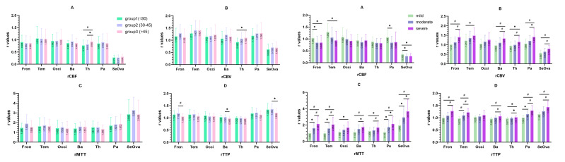
Results: Parameters in regional CBF (rCBF) of frontal, temporal lobe and basal ganglia in female was more superior than male. rCBF, regional MTT (rMTT) and regional TTP (rTTP) in adult MMD patients with haemorrhage were superior than the ischaemic. With the increase of age, significant difference could be seen in rCBF and rCBV of thalamus. However, with progress of arterial stenosis, significant difference could only be obsevrved in rCBV, rMTT and rTTP, whereas rCBF had no significant difference. For increase of moyamoya vessels, significant decrease of rCBF could be seen in temporal and parietal lobe. With the increase of compensatory artery numbers, no significant difference could be seen in rCBF parameters (p>0.05).

Conclusions: In adult MMD patients, age, gender and clinical type were potential risk factors for the change of cerebral perfusion. When arterial stenosis is worsened, moyamoya vessels could alter perfusion of temporal and parietal lobe, but not frontal lobe. Extracranial/intracranial compensatory arteries could maintain microcirculation stability in frontal lobe and basal ganglia, indicating that the protection from extracranial compensatory arteries, a theoretic base for surgery treatment if necessary.

Keywords: CT perfusion; angiography; blood flow; hemorrhage; stroke.


Conflict of interest statement

Competing interests: None declared.

Figures

Figure 1
Figure 1
ROI selection (similar to ASPECT scale) ROI 1 was vertebral artery (input artery) and ROI 2 was superior sagittal sinus (output vein). ROI 3 was set in cerebellum at level of dentatum nucleus. ROIs from 4 to 18 were as follows: superior frontal gyrus (ROI 4 and 14), inferior frontal gyrus (ROI 5 and 15), postocentral gyrus (ROI 6 and 16), supramarginal gyrus (ROI 7 and 17), occipital lobe (ROI 8 and 18), caudate nucleus (ROI 9), lenticular nucleus, external and internal capsule (ROI 10, 11 and 12) and thalamus (ROI 13). Anterior, central and posterior parts of centrum semiovale were established as ROI 19, 20 and 21. ROI, region of interest.
Figure 2
Figure 2
Univariate analysis of haemodynamic condition between gender and clinical type. Figure I and II were the relative value of rCBF, rCBV, rMTT and rTTP. The data of cerebellum at dentate nucleus on parameters of CBF, CBV, MTT and TTP were 61.51 mL/(min·100g), 2.74 mL/(min·100g), 2.86 s and 10.15 s, respectively. *P<0.05; #P<0.01. (Details could be seen in online supplementary material - figure 1). Ba, basal ganglia; CBF, cerebral blood flow; CBV, cerebral blood volume; Fron, frontal lobe; MTT, mean transit time; Occi, occipital lobe; Pa, parietal lobe; SeOva, centrum semiovale; Tem, temporal lobe; Th, thalamus; TTP, time to peak.
Figure 3
Figure 3
Univariate analysis of haemodynamic condition between age and degree of arterial stenosis. Figure III and IV were the relative value of rCBF, rCBV, rMTT and rTTP (A–D). The data of cerebellum at dentate nucleus on parameters of CBF, CBV, MTT and TTP were 61.51 mL/(min·100 g), 2.74 mL/(min·100 g), 2.86 s and 10.15 s respectively. *P<0.05; #P<0.01. (Details could be seen in online supplementary materials - figure 2). Ba, basal ganglia; CBF, cerebral blood flow; CBV, cerebral blood volume; Fron, frontal lobe; MTT, mean transit time; Occi, occipital lobe; Pa, parietal lobe; SeOva, centrum semiovale; Tem, temporal lobe; Th, thalamus; TTP, time to peak.
Figure 4
Figure 4
Univariate analysis of haemodynamic condition between compensatory artery numbers and moyamoya vesses. Figure V and VI were the relative value of rCBF, rCBV, rMTT and rTTP (A–D). The data of cerebellum at dentate nucleus on parameters of CBF, CBV, MTT and TTP were 61.51 mL/(min·100g), 2.74 mL/(min·100 g), 2.86 s and 10.15 s, respectively. *P<0.05; #P<0.01. (Details could be seen in online supplementary materials - figure 3). Ba, basal ganglia; CBF, cerebral blood flow; CBV, cerebral blood volume; Fron, frontal lobe; MTT, mean transit time; Occi, occipital lobe; Pa, parietal lobe; SeOva, centrum semiovale; Tem, temporal lobe; Th, thalamus; TTP, time to peak.
Figure 5
Figure 5
Multivariate analysis of haemodynamic condition between gender, clinical type, age, arterial stenosis, moyamoya vessels and compensatory artery numbers. All data were represented as relative value of rCBF, rCBV, rMTT and rTTP. Beta was represented as standardised coefficients. Ba, basal ganglia; CBF, cerebral blood flow; CBV, cerebral blood volume; Fron, frontal lobe; MTT, mean transit time; Occi, occipital lobe; Pa, parietal lobe; SeOva, centrum semiovale; Tem, temporal lobe; Th, thalamus; TTP, time to peak.

References

    1. Suzuki J, Kodama N. Moyamoya disease--a review. Stroke 1983;14:104–9. 10.1161/01.STR.14.1.104 - DOI - PubMed
    1. Amlie-Lefond C, Zaidat OO, Lew SM. Moyamoya disease in early infancy: case report and literature review. Pediatr Neurol 2011;44:299–302. 10.1016/j.pediatrneurol.2010.10.016 - DOI - PubMed
    1. Scott RM, Smith ER. Moyamoya disease and moyamoya syndrome. N Engl J Med 2009;360:1226–37. 10.1056/NEJMra0804622 - DOI - PubMed
    1. Sasagawa A, Mikami T, Hirano T, et al. . Characteristics of cerebral hemodynamics assessed by CT perfusion in moyamoya disease. J Clin Neurosci 2018;47:183–9. 10.1016/j.jocn.2017.09.020 - DOI - PubMed
    1. Han DH, Nam DH, Oh CW. Moyamoya disease in adults: characteristics of clinical presentation and outcome after encephalo-duro-arterio-synangiosis. Clin Neurol Neurosurg 1997;99 Suppl 2:S151–5. 10.1016/S0303-8467(97)00058-9 - DOI - PubMed

LinkOut - more resources