. 2020 Sep 22;117(38):23626-23635.
doi: 10.1073/pnas.2003228117. Epub 2020 Sep 3.

CHD7 and Runx1 interaction provides a braking mechanism for hematopoietic differentiation

Affiliations

CHD7 and Runx1 interaction provides a braking mechanism for hematopoietic differentiation

Jingmei Hsu et al. Proc Natl Acad Sci U S A. .

Erratum in

Abstract

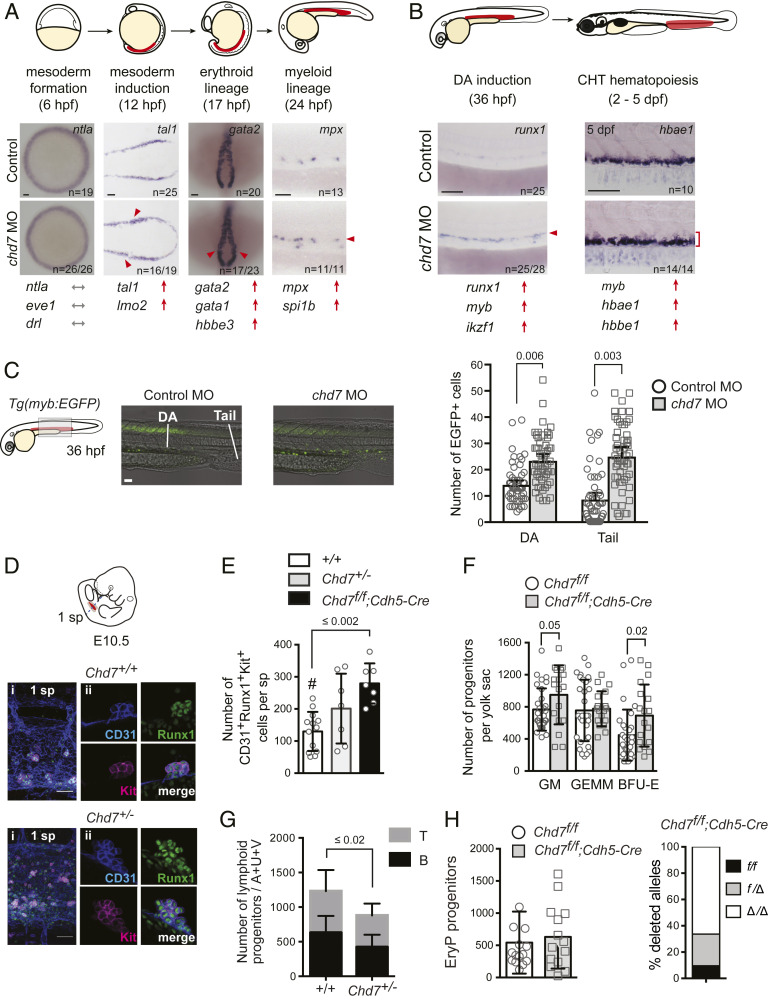
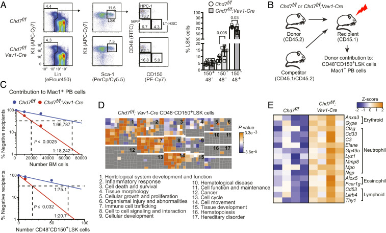
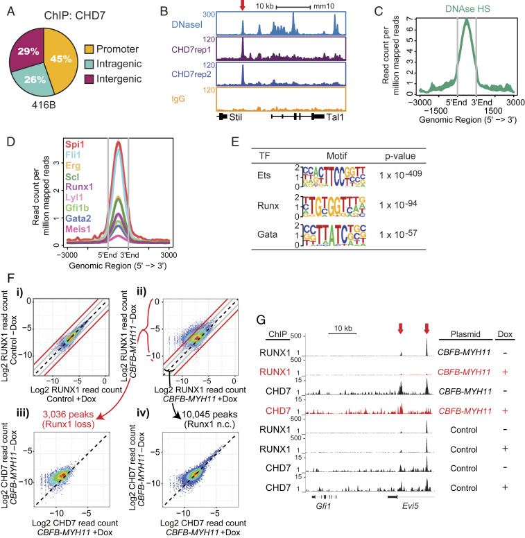
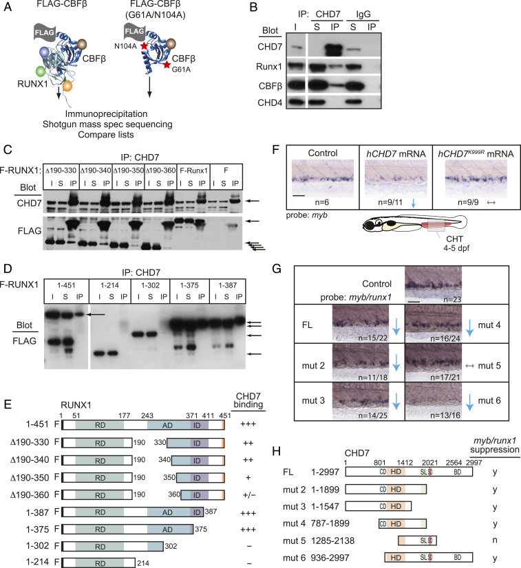
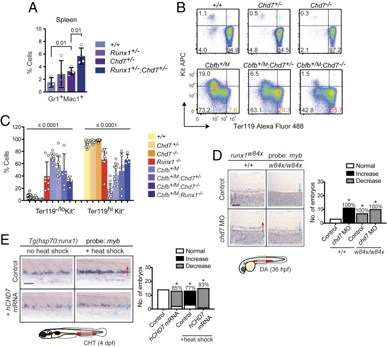
Hematopoietic stem and progenitor cell (HSPC) formation and lineage differentiation involve gene expression programs orchestrated by transcription factors and epigenetic regulators. Genetic disruption of the chromatin remodeler chromodomain-helicase-DNA-binding protein 7 (CHD7) expanded phenotypic HSPCs, erythroid, and myeloid lineages in zebrafish and mouse embryos. CHD7 acts to suppress hematopoietic differentiation. Binding motifs for RUNX and other hematopoietic transcription factors are enriched at sites occupied by CHD7, and decreased RUNX1 occupancy correlated with loss of CHD7 localization. CHD7 physically interacts with RUNX1 and suppresses RUNX1-induced expansion of HSPCs during development through modulation of RUNX1 activity. Consequently, the RUNX1:CHD7 axis provides proper timing and function of HSPCs as they emerge during hematopoietic development or mature in adults, representing a distinct and evolutionarily conserved control mechanism to ensure accurate hematopoietic lineage differentiation.

Keywords: CHD7; RUNX1; hematopoiesis.


Conflict of interest statement

Competing interest statement: L.I.Z. is founder and stockholder of Fate, Scholar Rock, and Camp4 therapeutics and a scientific advisor for Stemgent.

Figures

Fig. 1.
Fig. 1.
Chd7 negatively regulates embryonic hematopoiesis. (A) Chd7 knockdown increases expression of hematopoietic mesodermal precursor, primitive erythroid and myeloid, but not early mesoderm genes. Representative embryos for whole-mount in situ hybridization are shown, with additional genes shown in SI Appendix, Fig. S2. Regions of blood development are highlighted in red in the embryo schematic. Red arrows and arrowheads indicate an increase. Gray arrows indicate no change. (Scale bars, 50 μm.) Replicates: 2. (B) Chd7 knockdown increases expression of definitive HSPC and definitive myeloid and erythroid genes. Same symbols as in A. (C) Chd7 knockdown in Tg(myb:EGFP) embryos increases EGFP+ cells in the DA and tail region (Left), which is quantified in graph (Right) (n = 53 to 55). Representative embryos shown are from three independent replicates. (D) Chd7 deletion in mice increases Runx1+CD31+Kit+ hematopoietic clusters detected by confocal imaging of E10.5 Chd7+/+, Chd7+/−, and Chd7f/f; Cdh5-Cre AGM regions. Representative clusters shown. (i) One somite pair (sp) area. (ii) Individual cluster. (E) Quantification of data from D (n = 7 to 13). One-way ANOVA, Dunnett’s multiple comparison test; #, comparator. (F) Increased number of burst-forming unit–erythroid (BFU-E) and granulocyte/monocyte progenitors (colony-forming unit for granulocytes and macrophages) in E10.5 Chd7+/− yolk sacs (n = 8 to 14). GEMM, granulocyte/erythrocyte/monocyte/megakaryocyte progenitors. (G) Reduced number of lymphoid progenitors in E10.5 Chd7+/− embryos (n = 10 to 12). A+U+V: AGM, umbilical, and vitelline arteries. (H, Left) The number of erythroid progenitors (EryP) in the yolk sac of Chd7f/f;Cdh5-Cre embryos is not altered (n = 14 to 15). (H, Right) Both Chd7f alleles were deleted in 65% of the EryP colonies, and one allele was deleted in 27% of the colonies; thus Cdh5-Cre was active in the majority of EryPs or their precursors (n = colonies from 6 to 8 yolk sacs). All graphs show mean ± SD, unpaired two-tailed t test unless otherwise specified.
Fig. 2.
Fig. 2.
CHD7 regulates hematopoiesis cell autonomously. (A) Chd7 deficiency does not affect phenotypic LT-HSCs. Flow cytometry of LT-HSCs (CD48CD150+), MPPs (CD48CD150), and HPC-1s (CD48+CD150) from LinnegSca1+Kit+ (LSK) bone marrow populations (Left), which is quantified in bar graph (Right) (n = 6 to 7). Mean ± SD, unpaired two-tailed t test. (B) Schematic diagram of mouse limiting dilution transplantation experiments. (C) The frequency of functional CHD7-deficient LT-HSCs increased two-fold in whole BM (Left) and in purified CD48CD150+LSK cells (Right) when ≥1% donor contribution to Mac1+ PB was scored at 4 mo. LT-HSC frequency was calculated by ELDA (n = 7 to 14 recipients per dose). (D) Loss of CHD7 increases multilineage hematopoiesis. (Top) Functional categories for genes up-regulated in CHD7-deficient mouse LT-HSCs were enriched for hematopoietic-related functions by IPA. Clusters of individual functional gene annotations (squares) belonging to each category are labeled numerically. Replicates: 4. (E) Heatmap of representative lineage-specific genes up-regulated in CHD7-deficient CD48CD150+LSK cells.
Fig. 3.
Fig. 3.
CHD7 cooperates with hematopoietic transcription factors to regulate hematopoiesis. (A) CHD7-binding distribution in the murine 416B HPC cell line by ChIP-seq. Replicates: 2. (B) Gene track of CHD7 binding overlaps with DNaseI hypersensitive sites at the Tal1 gene. (C) Overlap of CHD7-binding and DNaseI hypersensitive sites. (D) Overlap of CHD7 binding and hematopoietic transcription factors. (E) CHD7-binding sites are highly enriched for Ets, Runx, and Gata motifs by HOMER motif analysis. (F) CHD7 binding is selectively lost from genomic regions where RUNX1 binding is attenuated by CBFB-MYH11. CBFB-MYH11 expression was induced in myeloid progenitor cells by doxycycline (Dox). RUNX1 occupancy in a (i) control clone and (ii) CBFB-MYH11-expressing clone. Loss of CHD7 occupancy is (iii) higher in regions of greater than four-fold RUNX1 occupancy loss and (iv) minimally changed in regions of less than two-fold RUNX1 occupancy loss. Diagonal black lines indicate no change (n.c.). Gray lines indicate two-fold change. Red lines indicate four-fold change. A replicate experiment is shown in SI Appendix, Fig. S5B. (G) Gene tracks showing loss of RUNX1 and CHD7 binding to Evi5 (red arrows) in Dox-induced CBFB-MYH11-expressing cells.
Fig. 4.
Fig. 4.
CHD7 interacts with Runx1 and restrains RUNX1 activity. (A) Scheme for identifying RUNX1-CBFβ–interacting proteins in a murine T-ALL cell line. FLAG-tagged CBFβ containing two amino acid substitutions (red stars) that decrease RUNX1 binding was used as a negative control. (B) CHD7 coimmunoprecipitates RUNX1-CBFβ but not CHD4 in murine T-ALL cells. I, input; S, depleted supernatant following immunoprecipitation; IP, immunoprecipitate. (C) Deletions impinging on the RUNX1 activation domain decrease the interaction between RUNX1 and CHD7. CHD7 was immunoprecipitated, and Western blots were probed with antibodies to CHD7 or FLAG. F-RUNX1, FLAG-RUNX1; ∆ refers to deleted amino acids illustrated schematically in panel E; F, vector expressing FLAG alone. Arrows indicate CHD7 (Top) or full-length and internally deleted RUNX1 proteins (Bottom). (D) C-terminal RUNX1 deletions. (E) Summary of RUNX1 mapping experiments. RD, DNA- and CBFβ-binding Runt domain; AD, transactivation domain; ID, inhibitory domain. (F) Expression of hCHD7 but not the catalytically dead mutant hCHD7K999R in zebrafish embryos reduces myb expression in the CHT by whole-mount in situ hybridization. Representative embryos are shown. Blue arrows indicate a decrease. Gray arrows indicate no change. (Scale bars, 50 μm.) Replicates: 2. (G) Mutation mapping of the hCHD7 domains shows that the ATPase/helicase domain is required to suppress myb and runx1 expression in the CHT. Same symbols as in F. (H) Summary of hCHD7 mapping experiments. FL, full length. CD, chromodomain. HD, ATPase/helicase domain. SL/SD/BD, SLIDE/SANT/BRK domains. y, yes; n, no. Quantification of results from F and G are in SI Appendix, Fig. S2B.
Fig. 5.
Fig. 5.
CHD7 interacts genetically with RUNX1 to regulate hematopoiesis. (A) Chd7 and Runx1 interact genetically to repress myelopoiesis in the spleen of adult mice by flow cytometric analysis (n = 4). (B) Restoration of primitive erythrocyte maturation with Chd7 deletion in CbfbM embryos with peripheral blood analysis by flow cytometry (n = 6 to 42). Representative plots are shown. Simplified genotypes are the following: Chd7+/-: Chd7+/f;β-actin-Cre, Chd7-/-: Chd7f/f;β-actin-Cre, Cbfb+/M: Cbfb+/MYH11;β-actin-Cre. (C) Chd7 or Runx1 deletion partially restores normal maturation of primitive erythrocytes in E10.5 embryos expressing the dominant neomorphic Cbfb-MYH11 allele (CbfbM). All values were significantly different as compared to Cbfb+/M. ANOVA and Dunnett’s multiple comparison test. (D) Expansion of myb+ HSPCs in chd7 morphant (MO) embryos is suppressed in runx1w84x mutants expressing truncated Runx1. Whole-mount in situ hybridization of representative embryos is shown, with phenotypic results quantified in bar graph (Right). *P < 0.01 by χ2 test. Red arrows, increase. Blue arrows, decrease. (Scale bar, 50 μm.) Replicates: 2. (E) Overexpression of hCHD7 suppresses the expansion of myb+ HSPCs caused by heat-shock–induced runx1 overexpression. Same descriptions and symbols as in D.

References

    1. Orkin S. H., Zon L. I., Hematopoiesis: An evolving paradigm for stem cell biology. Cell 132, 631–644 (2008). - PMC - PubMed
    1. Dzierzak E., Speck N. A., Of lineage and legacy: The development of mammalian hematopoietic stem cells. Nat. Immunol. 9, 129–136 (2008). - PMC - PubMed
    1. Huang H. T.et al. ., A network of epigenetic regulators guides developmental haematopoiesis in vivo. Nat. Cell Biol. 15, 1516–1525 (2013). - PMC - PubMed
    1. Sims J. K., Wade P. A., SnapShot: Chromatin remodeling: CHD. Cell 144, 626–626.e621 (2011). - PubMed
    1. Layman W. S., Hurd E. A., Martin D. M., Chromodomain proteins in development: Lessons from CHARGE syndrome. Clin. Genet. 78, 11–20 (2010). - PMC - PubMed

Publication types