Review
. 2020 Aug;53(2):207-218.
doi: 10.1055/s-0040-1716185. Epub 2020 Aug 29.

Reshaping the Lower Face Using Injectable Fillers

Affiliations
Review

Reshaping the Lower Face Using Injectable Fillers

André Braz et al. Indian J Plast Surg. 2020 Aug.

Abstract

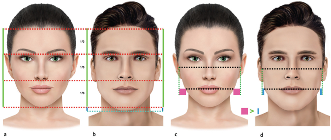
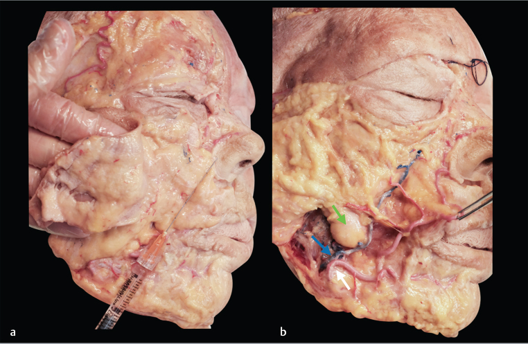
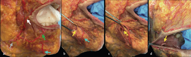
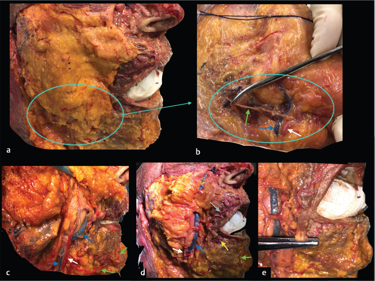
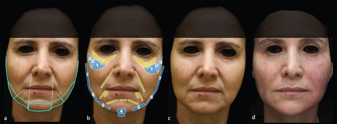
The lower third is very important for the pleasant appearance of the face. A well-contoured jawline is desirable in men and women, giving a perception of beauty and youth. It is also key to sexual dimorphism, defining masculine and feminine characteristics. The nonsurgical rejuvenation and beautification of the lower third of the face is becoming more frequent. Injectable fillers can reshape the jawline, lift soft tissues, and improve facial proportions, effectively improving the appearance of the area. It is paramount to understand the facial anatomy and perform a good facial assessment in order to propose a proper aesthetic treatment plan. The aesthetic goal of the rejuvenation approach is to redefine the mandibular angle and line. In young patients, beautification can be achieved through correction of constitutional deficit or enhancement of the contour of the face, improving the facial shape. It is very important to possess knowledge of facial anatomy and of the aging process in order to deliver effective and safe results. In this article, we discuss the anatomy of the lower third of the face, facial assessment, aging process, and treatments of the chin, prejowls and mandibular line and angle with injectable fillers. The authors' experience in the approach of this area is discussed.

Keywords: cosmetic therapies; dermal filler; facial rejuvenation; hyaluronic acid and calcium hydroxylapatite; lower face; nonsurgical.


Conflict of interest statement

Conflicts of Interest Dr. Eduardo reports personal fees from Galderma and personal fees from Underskin outside the submitted work. Dr. Braz reports personal fees from Allergan, personal fees from merz, personal fees from U.SK Under Skin, and personal fees from LÓreal outside the submitted work; in addition, Dr. Braz has a patent AB face technique pending.

Figures

Fig. 1
Fig. 1
Facial dimorphism. One of the aspects of beauty is the proportions among the three thirds of the face. In women, the height of the three thirds is similar ( a ), whereas in men, there is predominance of the lower third of the face ( b ) In women, there is a predominance in the middle third of the face: bizygomatic distance is wider than the bigonial distance ( c ) In men, theses distances are almost 1: 1.
Fig. 2
Fig. 2
Cadaveric dissection ( a ) The skin is folded back, showing the superficial fat compartments ( b ) Superficial fat and superficial musculoaponeurotic system (SMAS) are folded back ( c ) The superficial temporal artery, the parotid gland, the masseter muscle deeply, and the platysma muscle insertion in the mandible ( d ) The superficial temporal artery (red arrow), parotid gland (gray) masseter muscle (yellow arrow), platysma (light gray) facial vein (blue arrow), facial artery (white arrow), ascending to the face in the anterior masseter border, and buccal fat pad (green arrow).
Fig. 3
Fig. 3
The layers from superficial to deep: ( a ) Superficial subcutaneous fat ( b ) Deep mental fat and periosteum are exposed when the muscles are put laterally. ( c ) There are two possible depths of injection in the chin: blue colored gel injected in the superficial fat compartment and the green colored gel in the submuscular/supraperioteal plane.
Fig. 4
Fig. 4
The AB face technique approach to improve the structure of the face. It is important to restore the midface volume before approaching the lower face. Oblique view right side. ( a ) The marking of the areas to be injected. In yellow, the areas treated with hyaluronic acid (HA) filler Hydryalix gentle for refinement. The chin width is equivalent to the nose base width. ( b ) The filler was injected in the malar and zygomatic areas, from medial to lateral. The mental, prejowl, mandibular line, angle and ramus were treated. ( c ) Before and ( d ) after the treatment with Harmonyca and HA filler Hydryalix gentle (in yellow) with improved contours and proportion. The mandibular line was enhanced, softening the jowls, and keeping a feminine soft contour.
Fig. 5
Fig. 5
Cadaveric dissections. ( a ) The skin is folded back, showing the superficial fat compartment of the midface. ( b ) The superficial fat compartments were folded back, showing the facial vein (blue arrow) and facial artery (white arrow) that cross the mandible anterior to the masseter and are located in the deep fat compartments. Buccal fat pad (green arrow). The facial artery runs in the superficial subcutaneous in the peri alar region.
Fig. 6
Fig. 6
Cadaveric dissections. ( a ) Facial artery (gray arrow); inferior labial artery (green); superior labial artery (white); submental artery (blue). ( b ) The depressor anguli oris (DAO) muscle is put aside to show the mental artery emerging from the mental foramen (yellow arrow). The submental artery (blue arrow). ( c ) The mental artery (yellow arrow), deep to the DAO. ( d ) An intraoral view of the mental nerve (yellow arrow) that emerges from the mental foramen is located in line with the vertical axis of the second premolar tooth.
Fig. 7
Fig. 7
( a ) Superficial fat compartment. ( b ) The superficial fat compartment was folded back exposing the mandibular marginal branch of the facial nerve (green arrow) and the facial artery (white arrow) and vein (blue arrow). These structures are inferior to the mandibular border in this specimen, which is not the usual location for the mandibular marginal nerve anteriorly to the masseter, as discussed above. ( c ) Facial artery (white) and facial vein (blue) emerging in the face in anteriorly to the masseter border. The submental artery, cervical branch of the facial artery (green arrow) in the neck region, and in the chin. ( d ) Facial artery (white arrow) and facial vein (blue arrow). The inferior labial artery (yellow) and the superior labial artery (gray arrow), branches of the facial artery. The submental artery (green). ( e ) The depressor anguli oris (DAO) is clipped, showing the inferior labial artery deep to this muscle.
Fig. 8
Fig. 8
The AB face technique approach to improve the structure of the face. It is important to restore the midface volume before approaching the lower face, to achieve better results. Frontal view. ( a ) Show the authors’ marking of the areas to be injected. The chin width is equivalent to the nose base width. ( b ) The filler was injected in the malar and zygomatic areas, from medial to lateral. In the lower third, the mental, prejowl, mandibular line, angle and ramus were treated. ( c ) Before and ( d ) after the treatment with Harmonyca, with improved contours and proportion.
Fig. 9
Fig. 9
Approach to enhance the mandibular contour in a young woman. ( a ) The areas for treatment: chin, prejowl, mandibular line, angle and ramus. ( b ) Before the treatment and ( c ) after, showing an improvement in the mandibular contour, and enhancing the face and neck transition. ( d ) The second patient areas for treatment. As she has a recessed chin, the mental area was treated anteriorly and inferiorly, to project and elongate. ( e ) Before the treatment and ( f ) after the treatment with filler, an enhanced mandibular contour and projection of the chin.
Fig. 10
Fig. 10
( a ) and ( e ) show the treatment marking for the injection. Of note, the chin width is similar to the mouth width. ( b ) and ( f ) show the areas treated, the chin, prejowl, mandibular line, angle and ramus. ( c ) and ( g ) frontal and oblique views before the treatment. ( d ) and ( h ) frontal and oblique views, with an improved shape of the lower third. The bigonial distance is wider with a more defined jawline.
Fig. 11
Fig. 11
Clinical-anatomical correlation. ( a ) Skin. ( b ) The authors usually inject the filler in both planes, deep to the muscle and in the superficial subcutaneous. ( c ) The lateral approach to the chin, using cannula, which the authors find safer than using a needle, because of the lower risk of vascular adverse events.
Fig. 12
Fig. 12
Cadaveric dissection. ( a ) The superficial plane of injection in the chin. The cannula shape is seen below the skin. ( b ) The cannula in the superficial fat compartment. ( c ) The green colored gel place in the superficial fat.
Fig. 13
Fig. 13
Anatomical clinical correlation. ( a ) The superficial plane of injection in the chin. The cannula shape is seen below the skin. ( b ) The cannula in the superficial fat compartment. ( c ) The colored gel place in the superficial subcutaneous. ( d ) The injection of the prejowl region in the superficial subcutaneous plane
Fig. 14
Fig. 14
Anatomical clinical correlation. ( a ) The colored gel placed in the superficial fat (green arrow). The gel placed in the deep fat compartment (light blue arrow). Between these planes, the depressor anguli oris (DAO) (red arrow). ( b ) The injection of the prejowl region in the deep fat.
Fig. 15
Fig. 15
( a ) Cadaveric dissection showing the green colored gel placed in the superficial fat in the mandibular line, angle and ramus regions. The facial artery is shown in the deep fat, anterior to the masseter and ascending in the face. The superficial temporal artery. ( b ) The authors’ approach to the mandibular line. The entry point in the prejowl area. In pink is marked the anterior border of the masseter, where the facial artery crosses the mandible. ( c ) The authors’ approach to the mandibular line in another patient. The entry point in the prejowl area. In pink is marked the facial artery crossing the mandible. The second finger is palpating the mandibular angle, an important reference to the treatment.
Fig. 16
Fig. 16
( a ) Cadaveric dissection showing the green colored gel placed in the superficial fat in the mandibular line, angle and ramus regions. The cannula shows the mandibular ramus, preauricular and inferior zygomatic lateral area treatments. ( b and c ) Approach to these regions in two different patients.
Fig. 17
Fig. 17
( a ) The superficial temporal artery is shown. The filler must be placed in the superficial fat (above the superficial musculoaponeurotic system [SMAS]) in the preauricular and inferior zygomatic regions. ( b ) The facial transverse artery, the superficial temporal artery, and the cannula in the superficial fat compartments above these structures. ( c ) Facial transverse artery (light blue arrow) is located below the superficial fat compartment and SMAS. The injection plane is above this structure. The superficial temporal artery (dark blue arrow).
Fig. 18
Fig. 18
( a ) Simulating the treatment of the mandibular angle in the supraperiosteal plane. The masseter muscle (yellow), buccal fat pad (green), facial vein (blue) and facial artery (white arrow). ( b ) Red-colored gel in the mandibular angle. ( c ) Approach to the mandibular angle, injecting the filler in the supraperioteal plane, which is an option mainly when elongation of the mandibular line is desired.
Fig. 19
Fig. 19
( a ) Filler injection into the prejowl area with needle. Reflux maneuver showing blood in the syringe. ( b and c ) Injection in the chin. Reflux maneuver is paramount. Blood can be seen in the syringe, alerting to discontinue the injection to avoid placement of the product into a vessel. ( d ) Livedo reticularis in the chin area, a sign of skin ischemia, probably secondary to the commitment of the submental artery, cervical branch of the facial artery. This patient received injection in the chin in a position similar to the pictures ( b and c ) Of note, the possible positions of the submental artery, as shown in Figs. 6 7 .

References

    1. Shamban, A. The signature feature: a new concept in beauty. J Cosmet Dermatol 2019 (e-pub ahead of print). doi: https://doi.org/10.1111/jocd.12944 - PubMed
    1. Sykes J M, Fitzgerald R. Choosing the best procedure to augment the chin: is anything better than an implant? Facial Plast Surg. 2016;32(05):507–512. - PubMed
    1. Sands N B, Adamson P A. Global facial beauty: approaching a unified aesthetic ideal. Facial Plast Surg. 2014;30(02):93–100. - PubMed
    1. Goodman G J. The oval female facial shape- a study in beauty. Dermatol Surg. 2015;41(12):1375–1383. - PubMed
    1. Vazirnia A, Braz A, Fabi S G. Nonsurgical jawline rejuvenation using injectable fillers. J Cosmet Dermatol. 2019;00:1–8. - PubMed