

Electrical impedance tomography reveals pathophysiology of neonatal pneumothorax during NAVA
- PMID: 32884798
- PMCID: PMC7455437
- DOI: 10.1002/ccr3.2944
Electrical impedance tomography reveals pathophysiology of neonatal pneumothorax during NAVA
Abstract
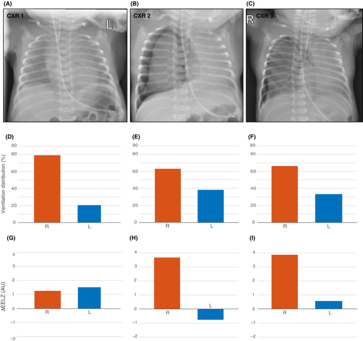
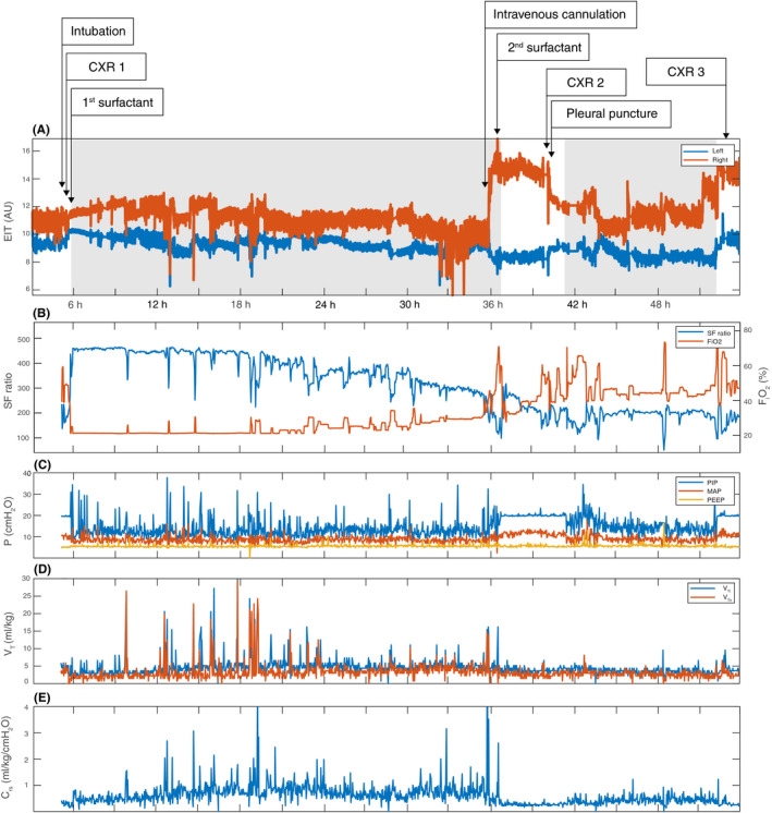
Pneumothorax is a potentially life-threatening complication of neonatal respiratory distress syndrome (RDS). We describe a case of a tension pneumothorax that occurred during neurally adjusted ventilatory assist (NAVA) in a preterm infant suffering from RDS. The infant was included in a multicenter study examining the role of electrical impedance tomography (EIT) in intensive care and therefore continuously monitored with this imaging method. The attending physicians were blinded for EIT findings but offline analysis revealed the potential of EIT to clarify the underlying cause of this complication, which in this case was heterogeneous lung disease resulting in uneven ventilation distribution. Instantaneous increase in end-expiratory lung impedance on the affected side was observed at time of the air leak. Real-time bedside availability of EIT data could have modified the treatment decisions made.
Keywords: electrical impedance tomography (EIT); neonate; neurally adjusted ventilatory assist; pneumothorax; respiratory distress syndrome (RDS).
© 2020 The Authors. Clinical Case Reports published by John Wiley & Sons Ltd.
Conflict of interest statement
The authors have no conflicts of interest to declare.
Figures
References
-
- Rahtu M, Frerichs I, Waldmann AD, et al. Early recognition of pneumothorax in neonatal RDS with electrical impedance tomography. Am J Respir Crit Care Med. 2019;200:1060‐1061. - PubMed
-
- Aly H, Massaro A, Acun C, Ozen M. Pneumothorax in the newborn: clinical presentation, risk factors and outcomes. J Matern Fetal Neonatal Med. 2014;27:402‐406. - PubMed
-
- Alander M, Peltoniemi O, Pokka T, Kontiokari T. Comparison of pressure‐, flow‐, and NAVA‐triggering in pediatric and neonatal ventilatory care. Pediatr Pulmonol. 2012;47:76‐83. - PubMed
-
- Brander L, Moerer O, Hedenstierna G, et al. Neural control of ventilation prevents both over‐distension and de‐recruitment of experimentally injured lungs. Respir Physiol Neurobiol. 2017;237:57‐67. - PubMed
-
- Miedema M, McCall KE, Perkins EJ, et al. First real‐time visualization of a spontaneous pneumothorax developing in a preterm lamb using electrical impedance tomography. Am J Respir Crit Care Med. 2016;194:116‐118. - PubMed
Publication types
LinkOut - more resources
Full Text Sources
Medical
