. 2020 Aug 20:2020:3413186.
doi: 10.1155/2020/3413186. eCollection 2020.

Kangtaizhi Granule Alleviated Nonalcoholic Fatty Liver Disease in High-Fat Diet-Fed Rats and HepG2 Cells via AMPK/mTOR Signaling Pathway

Affiliations

Kangtaizhi Granule Alleviated Nonalcoholic Fatty Liver Disease in High-Fat Diet-Fed Rats and HepG2 Cells via AMPK/mTOR Signaling Pathway

Jiaxin Zhang et al. J Immunol Res. .

Abstract

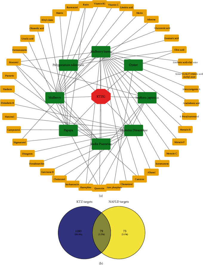
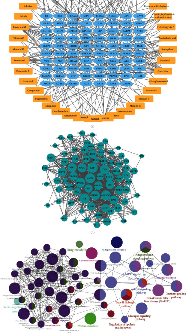
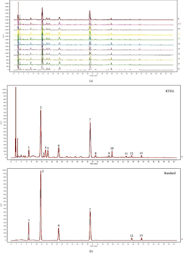
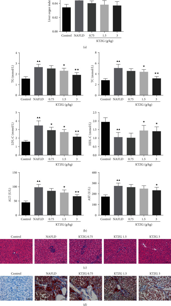
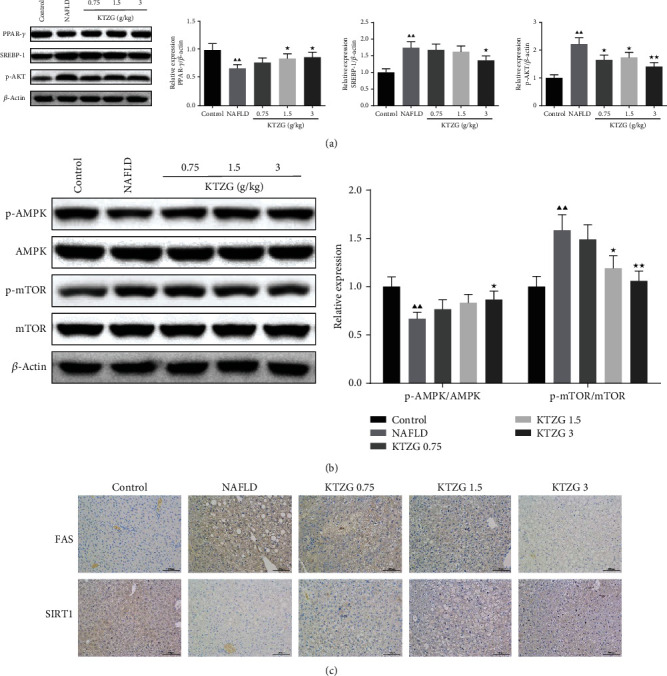
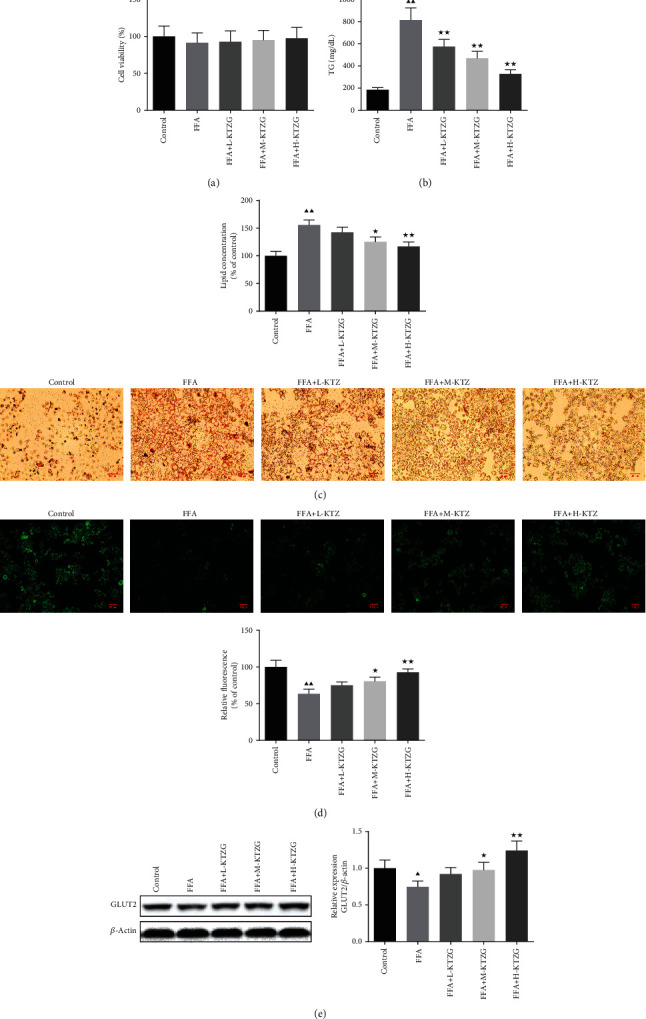
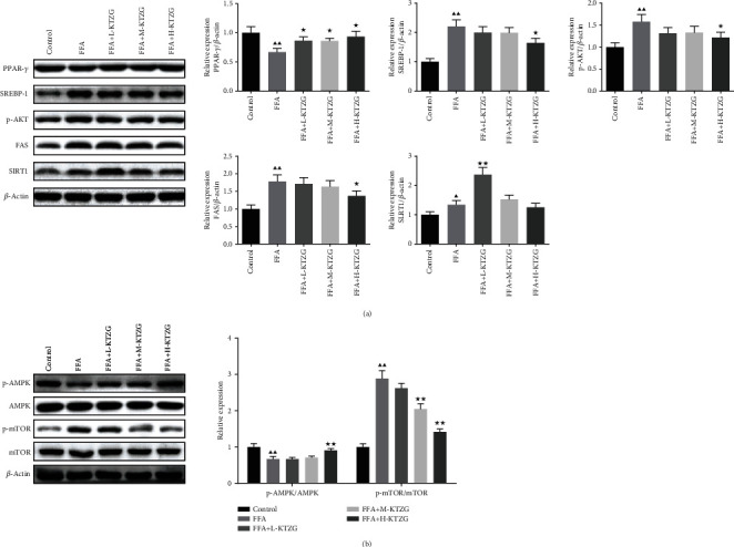
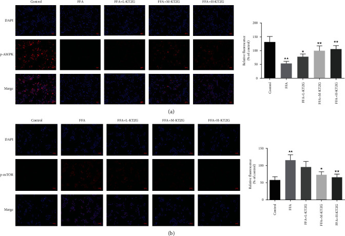
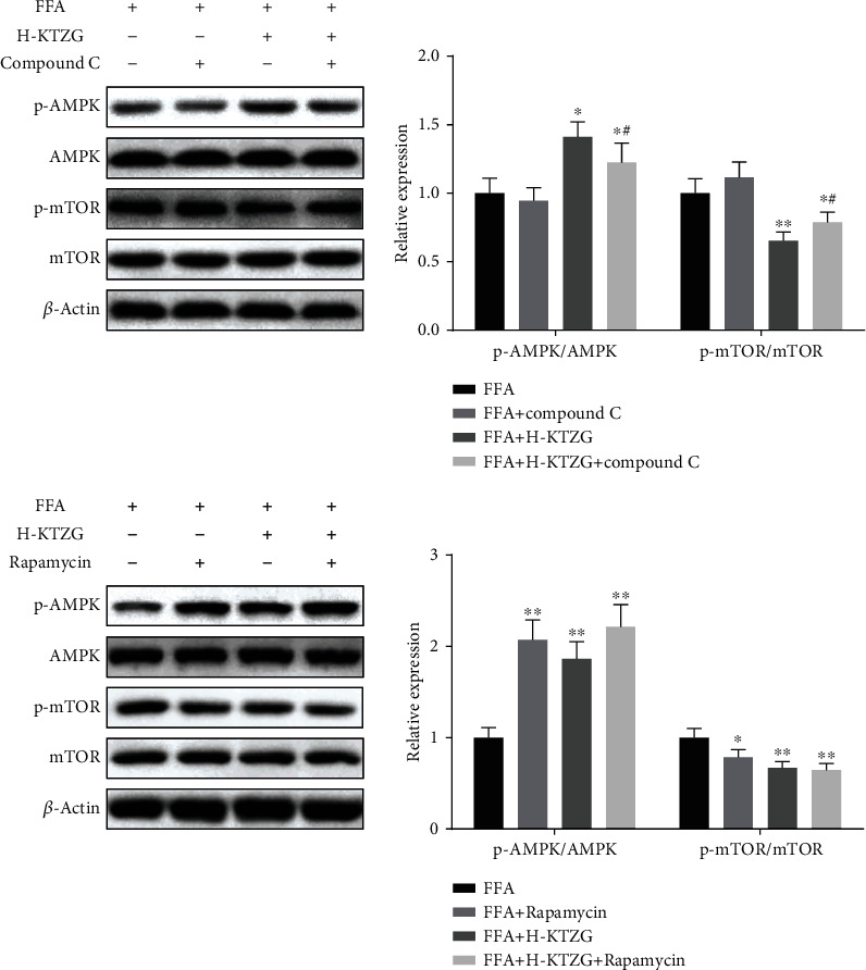
Kangtaizhi granule (KTZG) is a Chinese medicine compound prescription and has been proven to be effective in nonalcoholic fatty liver disease (NAFLD) treatment clinically. However, the underlying mechanisms under this efficacy are rather elusive. In the present study, network pharmacology and HPLC analysis were performed to identify the chemicals of KTZG and related target pathways for NAFLD treatment. Network pharmacology screened 42 compounds and 79 related targets related to NAFLD; HPLC analysis also confirmed six compounds in KTZG. Further experiments were also performed. In an in vivo study, SD rats were randomly divided into five groups: control (rats fed with normal diet), NAFLD (rats fed with high-fat diet), and KTZG 0.75, 1.5, and 3 groups (NAFLD rats treated with KTZG 0.75, 1.5, and 3 g/kg, respectively). Serum lipids were biochemically determined; hepatic steatosis and lipid accumulation were evaluated with HE and oil red O staining. In an in vitro study, HepG2 cells were incubated with 1 mM FFA to induce lipid accumulation with or without KTZG treatment. MTT assay, intracellular TG level, oil red O staining, and glucose uptake in cells were detected. Western blotting and immunohistochemical and immunofluorescence staining were also performed to determine the expression of lipid-related genes PPAR-γ, SREBP-1, p-AKT, FAS, and SIRT1 and genes in the AMPK/mTOR signaling pathway. In high-fat diet-fed rats, KTZG treatment significantly improved liver organ index and serum lipid contents of TG, TC, LDL-C, HDL-C, ALT, and AST significantly; HE and oil red O staining also showed that KTZG alleviated hepatic steatosis and liver lipid accumulation. In FFA-treated HepG2 cells, KTZG treatment decreased the intracellular TG levels, lipid accumulation, and attenuated glucose uptake significantly. More importantly, lipid-related genes PPAR-γ, SREBP-1, p-AKT, FAS, and SIRT1 expressions were ameliorated with KTZG treatment in high-fat diet-fed rats and FFA-induced HepG2 cells. The p-AMPK and p-mTOR expressions in the AMPK/mTOR signaling pathway were also modified with KTZG treatment in high-fat diet-fed rats and HepG2 cells. These results indicated that KTZG effectively ameliorated lipid accumulation and hepatic steatosis to prevent NAFLD in high-fat diet-fed rats and FFA-induced HepG2 cells, and this effect was associated with the AMPK/mTOR signaling pathway. Our results suggested that KTZG might be a potential therapeutic agent for the prevention of NAFLD.


Conflict of interest statement

The authors declare that there is no conflict of interest regarding the publication of this article.

Figures

Figure 1
Figure 1
KTZG-bioactive compounds and NAFLD-related target screening. (a) KTZG-bioactive compound network. (b) Screening of the protein-targeted KTZG for NAFLD treatment. KTZG: Kangtaizhi granule; NAFLD: nonalcoholic fatty liver disease.
Figure 2
Figure 2
Network pharmacology analysis of the KTZG for NAFLD treatment. (a) Compound-target network of KTZG. The network included 42 compounds and 79 proteins, forming 296 edges. (b) PPI network of the identified targets. Node size was positively associated with node degree. (c) KEGG pathway analysis of the targets.
Figure 3
Figure 3
HPLC chemical fingerprint of KTZG. (a) The reproducible HPLC chromatograms of HTT from 10 batches. (b) HPLC chemical fingerprint of KTZG with 6 peaks determined by comparing retention time with the standards: 3′-hydroxy puerarin (no. 1), puerarin (no. 2), daidzin (no. 6), rutin (no. 7), daidzein (no. 12), and quercetin (no. 13).
Figure 4
Figure 4
Effect of KTZG on lipid accumulation and hepatic steatosis in HFD-fed rats. (a) Liver organ index of the HFD-fed rat liver tissues treated with KTZG. (b) Serum content of TG, TC, LDL-c, HDL-c, ALT, and AST. (c) HE staining of liver tissues (magnification ×200). (d) Oil red O staining images of the HFD-fed rat liver tissues treated with KTZG (magnification ×200). HFD: high-fat diet. Compared to control group, P < 0.05, ▲▲P < 0.01; compared to NAFLD group, P < 0.05, ★★P < 0.01.
Figure 5
Figure 5
Effect of KTZG on PPAR-γ, SREBP-1, p-AKT, FAS, SIRT1, and AMPK/mTOR signaling pathway in HFD fed rats. (a) Western blotting for PPAR-γ, SREBP-1, and p-AKT in HFD-fed liver tissues treated with KTZG. (b) Western blotting for p-AMPK, AMPK, p-mTOR, and mTOR in HFD-fed liver tissues treated with KTZG. (c) Immunohistochemical of FAS and SIRT1 in liver tissues (magnification ×200). Compared to control group, P < 0.05, ▲▲P < 0.01; compared to NAFLD group, P < 0.05, ★★P < 0.01.
Figure 6
Figure 6
Effect of KTZG on lipid accumulation and hepatic steatosis in FFA-induced HepG2 cells. (a) MTT assay of the HepG2 cells. HepG2 cells were incubated in KZTG with FFA for 24 h. FFA: free fatty acids. (b) Intracellular TG levels in HepG2 cells treated with KTZG and FFA. (c) Oil red O staining images of HepG2 cells treated with KTZG and FFA (magnification ×200). (d) Effect of KTZG on the glucose uptake in FFA-induced HepG2 cells. (e) Western blotting for GLUT2 in HFD-fed liver tissues treated with KTZG. Compared to control group, P < 0.05, ▲▲P < 0.01; compared to FFA group, P < 0.05, ★★P < 0.01.
Figure 7
Figure 7
Effect of KTZG on PPAR-γ, SREBP-1, p-AKT, FAS, SIRT1, and AMPK/mTOR signaling pathway in FFA-induced HepG2 cells. (a) Western blotting for PPAR-γ, SREBP-1, and p-AKT. (b) Western blotting for p-AMPK, AMPK, p-mTOR, and mTOR. Compared to control group, P < 0.05, ▲▲P < 0.01; compared to FFA group, P < 0.05, ★★P < 0.01.
Figure 8
Figure 8
Immunofluorescence staining of p-AMPK (a) and p-mTOR (b) in FFA-induced HepG2 cells (magnification ×200). Compared to control group, P < 0.05, ▲▲P < 0.01; compared to FFA group, P < 0.05, ★★P < 0.01.
Figure 9
Figure 9
Regulation of KTZG on AMPK/mTOR signaling pathway in FFA-induced HepG2 cells. Compared to FFA group, P < 0.05, ★★P < 0.01; compared to FFA+compound C group, #P < 0.05, ##P < 0.01.

References

    1. Dang Y. Q., Hao S. J., Zhou W. J., Zhang L., Ji G. The traditional Chinese formulae Ling-gui-zhu-gan decoction alleviated non-alcoholic fatty liver disease via inhibiting PPP1R3C mediated molecules. BMC Complementary and Alternative Medicine. 2019;19(1):p. 8. doi: 10.1186/s12906-018-2424-1. - DOI - PMC - PubMed
    1. Cotter T. G., Rinella M. Nonalcoholic fatty liver disease 2020: the state of the disease. Gastroenterology. 2020;157(7):1851–1864. - PubMed
    1. Williams K. H., Shackel N. A., Gorrell M. D., McLennan S. V., Twigg S. M. Diabetes and nonalcoholic fatty liver disease: a pathogenic duo. Endocrine Reviews. 2013;34(1):84–129. doi: 10.1210/er.2012-1009. - DOI - PubMed
    1. Anstee Q. M., Mantovani A., Tilg H., Targher G. Risk of cardiomyopathy and cardiac arrhythmias in patients with nonalcoholic fatty liver disease. Nature Reviews. Gastroenterology & Hepatology. 2018;15(7):425–439. doi: 10.1038/s41575-018-0010-0. - DOI - PubMed
    1. Singal A. G., Manjunath H., Yopp A. C., et al. The effect of PNPLA3 on fibrosis progression and development of hepatocellular carcinoma: a meta-analysis. The American Journal of Gastroenterology. 2014;109(3):325–334. doi: 10.1038/ajg.2013.476. - DOI - PMC - PubMed

MeSH terms