

Macrophage polarization in peri-implantitis lesions
- PMID: 32886246
- PMCID: PMC7966129
- DOI: 10.1007/s00784-020-03556-2
Macrophage polarization in peri-implantitis lesions
Abstract
Objectives: To immunohistochemically characterize and correlate macrophage M1/M2 polarization status with disease severity at peri-implantitis sites.
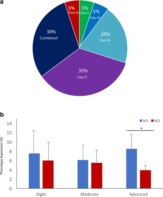
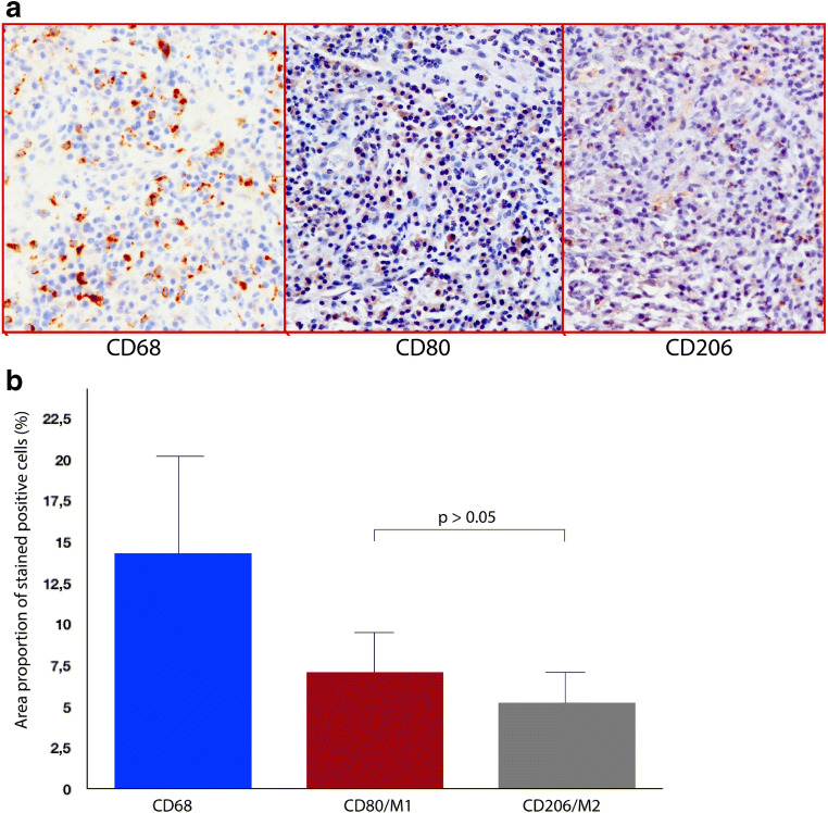
Materials and methods: A total of twenty patients (n = 20 implants) diagnosed with peri-implantitis (i.e., bleeding on probing with or without suppuration, probing depths ≥ 6 mm, and radiographic marginal bone loss ≥ 3 mm) were included. The severity of peri-implantitis was classified according to established criteria (i.e., slight, moderate, and advanced). Granulation tissue biopsies were obtained during surgical therapy and prepared for immunohistological assessment and macrophage polarization characterization. Macrophages, M1, and M2 phenotypes were identified through immunohistochemical markers (i.e., CD68, CD80, and CD206) and quantified through histomorphometrical analyses.
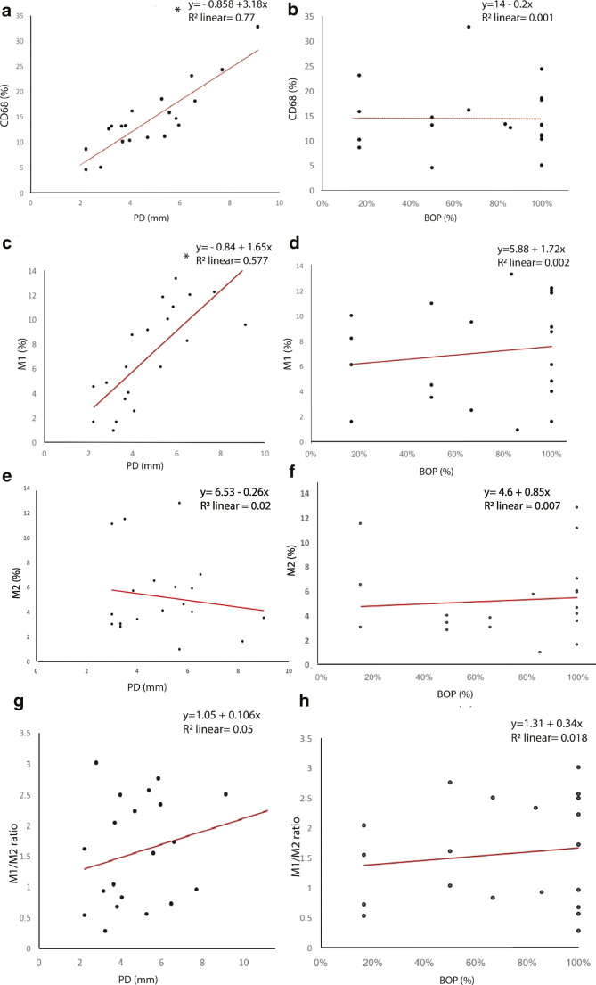
Results: Macrophages exhibiting a positive CD68 expression occupied a mean proportion of 14.36% (95% CI 11.4-17.2) of the inflammatory connective tissue (ICT) area. Positive M1 (CD80) and M2 (CD206) macrophages occupied a mean value of 7.07% (95% CI 5.9-9.4) and 5.22% (95% CI 3.8-6.6) of the ICT, respectively. The mean M1/M2 ratio was 1.56 (95% CI 1-12-1.9). Advanced peri-implantitis cases expressed a significantly higher M1 (%) when compared with M2 (%) expression. There was a significant correlation between CD68 (%) and M1 (%) expression and probing depth (PD) values.
Conclusion: The present immunohistochemical analysis suggests that macrophages constitute a considerable proportion of the inflammatory cellular composition at peri-implantitis sites, revealing a significant higher expression for M1 inflammatory phenotype at advanced peri-implantitis sites, which could possibly play a critical role in disease progression.
Clinical relevance: Macrophages have critical functions to establish homeostasis and disease. Bacteria might induce oral dysbiosis unbalancing the host's immunological response and triggering inflammation around dental implants. M1/M2 status could possibly reveal peri-implantitis' underlying pathogenesis.
Keywords: Biopsy; Dental implant; Immunohistochemistry; Macrophage polarization; Peri-implantitis.
Conflict of interest statement
The authors declare that they have no conflict of interest.
Figures
References
-
- Stöger JL, Gijbels MJJ, Van Der Velden S et al (2012) Distribution of macrophage polarization markers in human atherosclerosis. 10.1016/j.atherosclerosis.2012.09.013 - PubMed
-
- Hirata Y, Tabata M, Kurobe H et al (2011) Coronary atherosclerosis is associated with macrophage polarization in epicardial adipose tissue. 10.1016/j.jacc.2011.01.048 - PubMed
MeSH terms
Substances
Grants and funding
LinkOut - more resources
Full Text Sources
