Review
. 2020 Nov 15;3(1):59.
doi: 10.1186/s42155-020-00150-1.

Managing systemic venous occlusions in children

Affiliations
Review

Managing systemic venous occlusions in children

Anne E Gill et al. CVIR Endovasc. .

Abstract

Pediatric venous disease is increasing in incidence in both inpatient and outpatient populations. The widespread use of central venous access devices as well as the rising incidence of thromboembolic events in pediatrics is leading to more systemic venous occlusions in both the central and peripheral veins. This review focuses on the etiology, presentation, workup, and general technical considerations of recanalization as well as procedural complications related to pediatric systemic venous occlusive disease. The potential role for pediatric interventional radiology guided treatments will be discussed in detail.

Keywords: Endovascular stenting; Endovascular venous reconstruction; Inferior vena cava; Pediatrics; Superior vena cava; Systemic venous occlusion; Thromboembolism.


Conflict of interest statement

The authors declare that they have no competing interests.

Figures

Fig. 1
Fig. 1
5-year-old female with history of May-Thurner syndrome presenting with acute left lower extremity DVT and skin changes characteristic of phlegmasia cerulea dolens. [Image adapted from Kuo I, Smith J, Abou-Zamzam AM Jr. A multimodal therapeutic approach to phlegmasia cerulea dolens in a pediatric patient. J Vasc Surg. 2011. Jan;53(1):212–5. doi: 10.1016/j.jvs.2010.07.067. Epub 2010 Sep 26. PubMed PMID: 20875715]
Fig. 2
Fig. 2
a MRV of an 18 y/o M with chronic right lower extremity swelling and DVT demonstrates numerous venous collaterals but no central draining vein beyond the level of the iliac veins. This delayed, post-contrast MIP image shows the numerous venous collaterals in the pelvis and along the spine. b MRV of a 16 y/o F with acute onset of left lower extremity DVT shows patency of the right iliac veins draining into the IVC but no opacification of the left iliac veins. The overriding right common iliac artery compresses the left common iliac vein leading to the delayed venous drainage of the extremity and eventual thrombus formation. c CT of a 4 y/o M with chronic occlusion of the IVC (dashed arrow), multifocal calcified thrombi in the IVC (solid arrow), and fresh acute clot in the right common iliac vein (dotted arrow). A CT exam in this child allowed for an accurate, high resolution exam for deep venous recanalization planning without having to put the child under general anesthesia to obtain the images
Fig. 3
Fig. 3
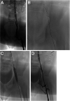
a 8 y/o M with history of left lower extremity DVT 1 month after ECMO cannulation for respiratory and cardiac failure. The left femoral vein was used for venous access for ECMO cannula. Venogram demonstrates short segment occlusion of the left external iliac with patent left common femoral, common iliac vein, and IVC. b A 0.035″ stiff hydrophilic wire was passed across the occlusion and through the native vessel lumen. c Balloon angioplasty was performed with noncompliant balloon; no appreciable waist present with balloon inflation. d Final venogram demonstrates a patent channel with persistent collateral vessels and no extravasation or vessel injury
Fig. 4
Fig. 4
14 y/o M with chronic venous occlusion of the right lower extremity. Solid arrow marks the venous cap: the antegrade entrance to the occluded vessel lumen. Dashed arrow marks the wire looped within the central lumen of the patent portion of the peripheral vessel
Fig. 5
Fig. 5
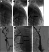
18 y/o M with chronic right lower extremity swelling and DVT presents for planning venogram after MRI demonstrated IVC agenesis from the level of the external iliac to the suprarenal IVC. a Initial venogram from the right groin shows numerous diminutive collateral vessels (dotted arrow). A wire was able to be passed from the left groin through collateral vessels (dashed arrow) to the suprarenal IVC (solid arrow). b Wires and catheters were eventually able to be passed through retroperitoneal collaterals until the bilateral accesses reached the suprarenal IVC (right groin access marked by dotted arrow, left groin access marked by dashed arrow, IVC solid arrow). c IVC reconstruction with stent complex placed from bilateral groins to the suprarenal IVC. d Final venogram through stent complex showed excellent outflow with no intrastent stenosis or delay in transit of contrast. e Follow up venogram at POD 45, demonstrated continued excellent outflow with no intrastent stenosis or occlusion. Patient’s symptoms of right lower extremity swelling was completely resolved. f Follow up venogram 5 months following stent placement, patient had begun to have difficulty with anticoagulation compliance and exhibited symptoms of leg swelling. Unable to pass a wire or catheter beyond the area marked with the solid arrow. g Accompanying venogram depicting complete occlusion of the right stent complex with chronic thrombus and numerous collateral vessels draining the right lower extremity (Note, patient is positioned prone). On IVUS, there was kinking of the stent complex near the femoral head which likely was at least partially contributing to the stent complex failure
Fig. 6
Fig. 6
5 y/o M with end stage renal disease requiring hemodialysis with very limited central venous access (left internal jugular occluded and left femoral vein occluded). His hemodialysis catheter was inadvertently pulled out and central venous access lost. a Initial venogram from the right neck demonstrates numerous cervical collateral vessels (solid arrow) with no opacification of the right internal jugular vein. Collateral vessels eventually drain into the patent mid SVC and azygous vein (dashed arrow). b 22G 10cm Chiba needle passed behind the clavicle (solid arrow) part way with ultrasound guidance and residual guidance with fluoroscopy. Patent SVC target is marked with angled catheter and wire (dashed arrow). c Wire is then passed from the neck to the IVC and safety wire from the groin is retracted. Eventually the wire from the neck was snared and “flossed” access was achieved. d Post recanalization venogram demonstrates patent but slightly narrowed SVC with limited cervical collateral filling. e Hemodialysis catheter placement with tip terminating in the right atrium and tunnel created over the shoulder to reduce the risk of inadvertent catheter removal in the future
Fig. 7
Fig. 7
4 y/o M with end stage renal disease requiring hemodialysis access and complete occlusion of the bilateral internal jugular veins and bilateral brachiocephalic veins. Hemodialysis access was from the right groin but he developed thrombus in that vessel and there was concern he would lose his ability for transplant consideration; thus access from the neck was desired. a Wire and sheath access from the right groin into the patent portion of the SVC. Snare and sheath access from a collateral vessel in the neck. Wire is also noted on the left periphery of the image marking the subclavian vein into the azygous vein. b A 22G 65 cm Chiba needle (solid arrow) was passed from the right groin sheath into the snare (dashed arrow) from the right neck venous access. c Inner obturator of needle was removed and replaced with a 0.018″ hydrophilic wire. Wire (solid arrow) was advanced centrally and snare tightened on wire (dashed arrow) while needle was retracted. d Sheath was advanced through the occlusion (dashed arrow) and venogram performed. Demonstrated numerous collateral venous vessels (upper solid arrow) with patent azygous vein (lower solid arrow) but no opacification of the occluded SVC. e Stent complex: balloon mounted Palmaz stent (Cordis Baar, Switzerland) deployed at the level of the complete occlusion (two solid arrows marking the cranial and caudal extent of the stent) and a self-expanding, Zilver 14 mm stent (Cook Bloomington, IN) deployed within the Palmaz stent. The stent complex connected the patent vessel lumens (two dashed arrows marking the cranial and caudal extent of the stent). Center of stent complex only ballooned to 10 mm due to size of patient native vasculature with intent of subsequent procedures to increase stent diameter as the patient grows. Dotted arrow marks the wire in the subclavian vein. f Final venogram status post recanalization, stenting, and balloon angioplasty. Contrast readily fills the right atrium with no extravasation present

References

    1. Al-Hakim RA, Kaufman JA, Farsad K. Iliac vein stent placement: acute Venographic changes and relevance to venous biomechanics. J Vasc Interv Radiol. 2018;29:1023–1027. doi: 10.1016/j.jvir.2018.02.031. - DOI - PubMed
    1. Andrew M, David M, Adams M, et al. Venous thromboembolic complications (VTE) in children: first analyses of the Canadian registry of VTE. Blood. 1994;83:1251–1257. doi: 10.1182/blood.V83.5.1251.1251. - DOI - PubMed
    1. Bass JE, Redwine MD, Kramer LA, et al. Spectrum of Congenital Anomalies of the Inferior Vena Cava: Cross-sectional Imaging Findings: (CME available in print version and on RSNA Link) RadioGraphics. 2000;20:639–652. doi: 10.1148/radiographics.20.3.g00ma09639. - DOI - PubMed
    1. Farsad K, Fuss C, Kolbeck KJ, et al. Transjugular intrahepatic Portosystemic shunt creation using intravascular ultrasound guidance. J Vasc Interv Radiol. 2012;23:1594–1602. doi: 10.1016/j.jvir.2012.08.023. - DOI - PubMed
    1. Frazer JR, Ing FF. Stenting of stenotic or occluded iliofemoral veins, superior and inferior vena cavae in children with congenital heart disease: acute results and intermediate follow up. Catheter Cardiovasc Interv. 2009;73:181–188. doi: 10.1002/ccd.21790. - DOI - PubMed

LinkOut - more resources