. 2020 Sep 4;15(9):e0238750.
doi: 10.1371/journal.pone.0238750. eCollection 2020.

Corneal dysfunction precedes the onset of hyperglycemia in a mouse model of diet-induced obesity

Affiliations

Corneal dysfunction precedes the onset of hyperglycemia in a mouse model of diet-induced obesity

Aubrey Hargrave et al. PLoS One. .

Abstract

Purpose: The purpose of this study was to use a mouse model of diet-induced obesity to determine if corneal dysfunction begins prior to the onset of sustained hyperglycemia and if the dysfunction is ameliorated by diet reversal.

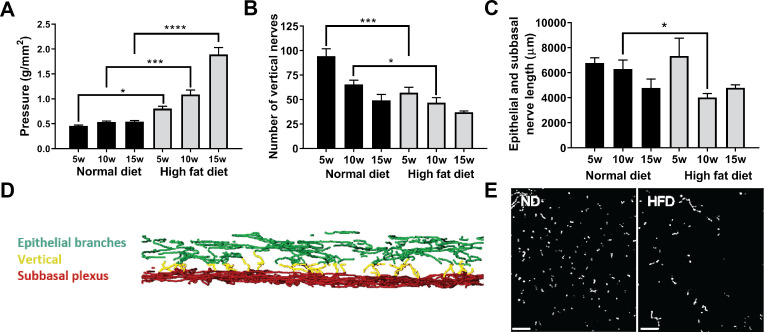
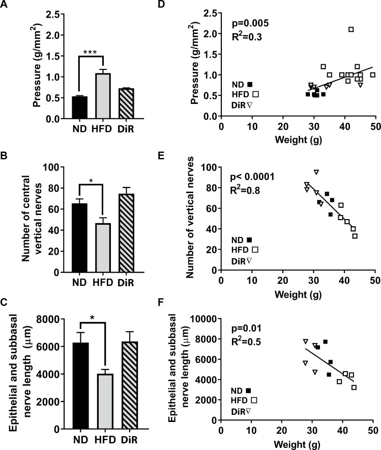
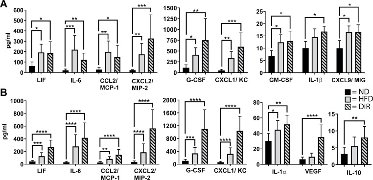
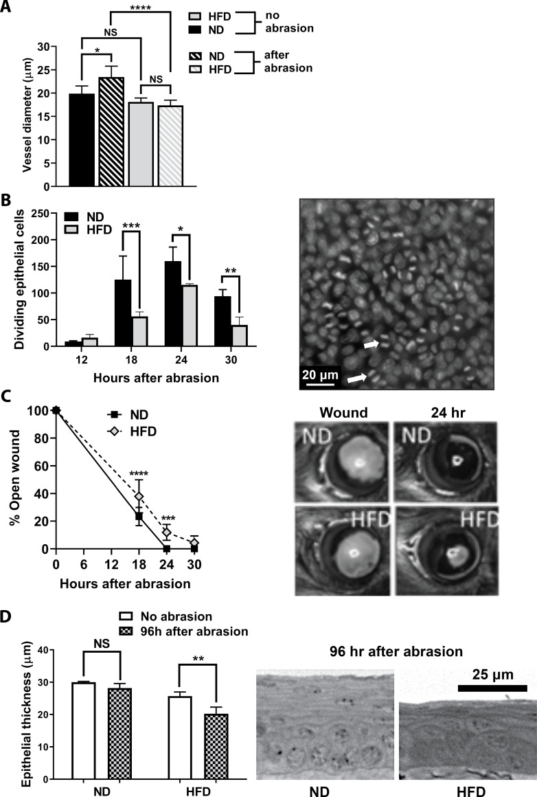
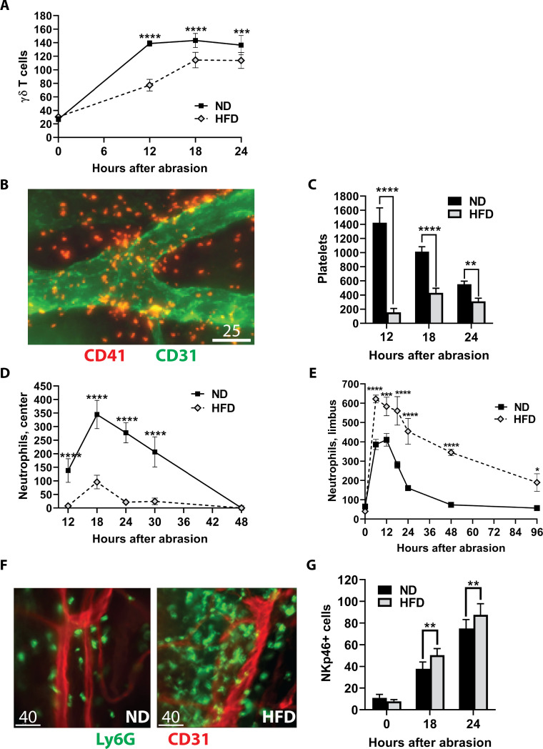
Methods: Six-week-old male C57BL/6 mice were fed a high fat diet (HFD) or a normal diet (ND) for 5-15 weeks. Diet reversal (DiR) mice were fed a HFD for 5 weeks, followed by a ND for 5 or 10 weeks. Corneal sensitivity was determined using aesthesiometry. Corneal cytokine expression was analyzed using a 32-plex Luminex assay. Excised corneas were prepared for immunofluorescence microscopy to evaluate diet-induced changes and wound healing. For wounding studies, mice were fed a HFD or a ND for 10 days prior to receiving a central 2mm corneal abrasion.

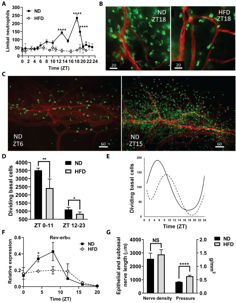
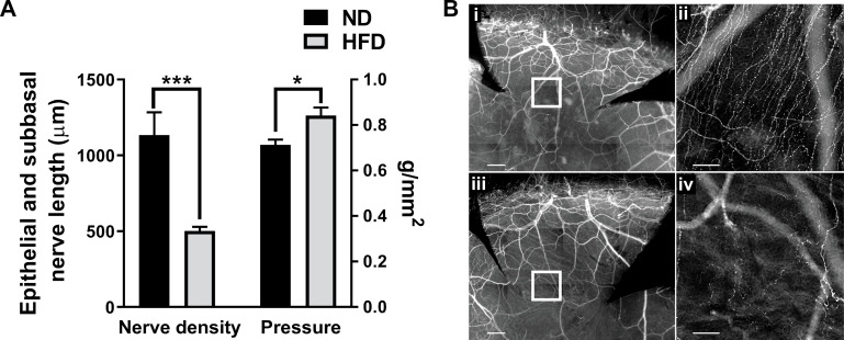
Results: After 10 days of HFD consumption, corneal sensitivity declined. By 10 weeks, expression of corneal inflammatory mediators increased and nerve density declined. While diet reversal restored nerve density and sensitivity, the corneas remained in a heightened inflammatory state. After 10 days on the HFD, corneal circadian rhythms (limbal neutrophil accumulation, epithelial cell division and Rev-erbα expression) were blunted. Similarly, leukocyte recruitment after wounding was dysregulated and accompanied by delays in wound closure and nerve recovery.

Conclusion: In the mouse, obesogenic diet consumption results in corneal dysfunction that precedes the onset of sustained hyperglycemia. Diet reversal only partially ameliorated this dysfunction, suggesting a HFD diet may have a lasting negative impact on corneal health that is resistant to dietary therapeutic intervention.


Conflict of interest statement

The authors have declared that no competing interests exist.

Figures

Fig 1
Fig 1. Changes in corneal nerves and sensitivity with HFD.
C57BL/6 male mice were fed the ND or HFD for up to 15 weeks. (A) Cochet-Bonnett aesthesiometry was used to assess corneal sensitivity to touch before corneas were collected at 5, 10 or 15 weeks of feeding for corneal nerve analysis. Sensitivity is shown as the pressure necessary to induce a blink response (g/mm2) (n = 28 ND, n = 35 HFD), (B) Morphometric analysis of the number of vertical nerves was determined by examining full thickness z-stacks of the epithelium (n = 12 ND, n = 13 HFD), (C) Analysis of the total length (μm) of epithelial nerves (epithelial branches and subbasal plexus) (n = 10 ND, n = 11 HFD), (D) Three-dimensional reconstruction of the epithelial nerves showing the subbasal plexus (red), epithelial nerve branches (green) and the vertical nerves (yellow) extending between them, and (E) en face projected image of the region of the epithelium containing vertical nerves (anti-β tubulin III, scale bar = 30 μm). Data shown as means ± SEM. *p<0.05, ***p<0.001, ****p<0.0001.
Fig 2
Fig 2. Changes in corneal nerves and sensitivity with diet reversal.
C57BL/6 male mice were fed the ND or HFD for 10 weeks, and diet reversal (DiR) mice were fed the HFD for 5 weeks followed by the ND for 5 weeks. (A) Cochet-Bonnett aesthesiometry was used to assess corneal sensitivity to touch (g/mm2) (n = 10 ND, n = 13 HFD, n = 12 DiR). Corneas were analyzed for corneal nerve density, including (B) number of vertical nerves (n = 4 ND, n = 5 HFD, n = 6 DiR) or (C) total length in μm of epithelial branches and the subbasal plexus (n = 4 per group). Data shown as means ± SEM. *p<0.05, ***p<0.001. Pearson correlations of these measures with body weight for ND, HFD and DiR at 10 weeks are also plotted (D-F).
Fig 3
Fig 3. Inflammatory protein expression in corneas.
C57BL/6 male mice were fed the ND or HFD for 10 weeks, and diet reversal (DiR) mice were fed the HFD for 5 weeks followed by the ND for 5 weeks. Corneas (n = 6 per group) were excised and cultured for 6h in vitro, and the (A) corneal extracts and (B) culture supernatant were analyzed for chemokines, cytokines and growth factors. Data shown as means ± SD. *p<0.05, **p<0.01, ***p<0.001, ****p<0.0001.
Fig 4
Fig 4. Corneal changes after 10 days on the high fat diet.
(A) Neutrophils were counted in four 40X microscope fields of view in extravascular limbal tissue at intervals over a 24h cycle (n = 4 corneas per group at each time point). (B) Representative micrographs of neutrophils in the limbus at ZT18, comparing limbal regions of mice fed a ND and a HFD; neutrophils (green), limbal blood vessels (red), scale bar = 20 μm. (C) Representative micrographs of neutrophils around the limbal vascular network at ZT6 and ZT15 in corneas from mice on ND; neutrophils (green), limbal blood vessels (red), scale bar = 60 μm. (D) The average of the sum of mitotic cells observed in the basal epithelium in nine perpendicular (horizontal and vertical) 40X microscopic fields across the cornea from limbus to limbus between ZT0-ZT11 and ZT12-ZT23 (n = 4 corneas per group collected every hour for 24h). (E) Changes in the number of mitotic cells in corneal epithelial cells over a 24h cycle. A polynomial function was used to fit the curve for the dividing cell number (the average of the sum of mitotic cells counted in nine vertical and horizontal 40X fields at each time point) with n = 4 corneas collected every hour for 24h (ZT scale). Solid line = ND, dotted line = HFD. (F) Expression of clock gene Rev-erbα over a 24h cycle (n = 2 independent pooled samples from 6–8 mice at each time point). (G) Nerve density (total axon length (μm) of the epithelial branches and subbasal plexus) in the corneal epithelium of ND mice and HFD mice (n = 4 per group). Corneal sensitivity, measured as the tactile pressure (g/mm2) required to elicit a blink, in mice fed the HFD for 10 days compared to ND fed mice (n = 6 per group). Data shown as means ± SD (A-E) and SEM (F, G). *p<0.05, **p<0.01, ****p<0.0001.
Fig 5
Fig 5. Inflammatory response in the cornea after epithelial abrasion in ND and HFD fed mice.
(A) Venule diameters in the corneal limbus of mice fed a ND or a HFD before and 24h after receiving a corneal abrasion (n = 5 per group). (B) Dividing basal epithelial cells counted in nine perpendicular 40X fields of view across the cornea (n = 4 per group). Representative image of epithelial cell division in the basal epithelium at 18h after wounding (scale bar = 20 μm). Arrows indicate two examples of mitosis. (C) Wound closure was assessed by fluorescein staining of the open wound at different times after abrasion (n = 8 per group). Representative image of the epithelial wound immediately following abrasion (left) and 24h after abrasion (right) in ND and HFD mice. (D) Epithelial thickness (μm) was determined at 96h after epithelial abrasion in fixed and sectioned plastic embedded corneas (n = 4–5 per group). Representative images shown in the cross sections (scale bar = 25 μm). Data represented as means ± SD (A-C) and SEM (D). *p<0.05, **p<0.01, ***p<0.001, ****p<0.0001.
Fig 6
Fig 6. Effect of the HFD on corneal responses to central epithelial abrasion.
Corneas from ND and HFD fed mice were analyzed after epithelial abrasion. (A) γδ T cells were counted in the epithelium and stroma across the cornea and plotted at 0, 6, 12, and 18h after epithelial abrasion (n = 6 per group). (B) Representative micrograph of extravascular platelets (red) around the limbal vascular network (green) at 12h after injury in a mouse fed a ND. Scale bar = 25 μm. (C) The sum of platelets in eight random non-overlapping 40X fields of view in the corneal limbus determined at 12, 18 and 24h after corneal abrasion (n = 4 at each time point). (D) Kinetics of neutrophil influx into the central abraded area of the cornea (counts from four 40X fields in the center of the cornea, n = 8 at each time point) are plotted at 12, 18, 24, 30, and 48h after abrasion. (E) Kinetics of neutrophil influx into the corneal limbus (counts from eight 40X fields in the limbus, n = 4–8 at each time point) prior to wounding (0h) and at 6, 12, 18, 24, 48, and 96h after abrasion. (F) Representative micrographs of neutrophil (green) influx around the limbal vascular (red) network at 48h after injury. Scale bar = 40 μm. (G) Limbal stromal NK cell counts (determined from nine vertical and horizontal 40X fields across the cornea, n = 8 per group) without wounding (0h), and at 18 and 24h after epithelial abrasion. Data shown as means ± SD. *p<0.05, **p<0.01, ***p<0.001, ****p<0.0001.
Fig 7
Fig 7. Healing nerves after corneal epithelial abrasion.
Corneas from ND and HFD fed mice were analyzed after epithelial abrasion. (A) At 96h post-injury, nerve density (total axon length (μm) of the epithelial branches and subbasal plexus) recovery (left) in 10d HFD fed mice compared with those on the ND (n = 5 per group). Cochet-Bonnett aesthesiometry was used to assess corneal sensitivity to touch (g/mm2) at 96h post-injury (right) in 10d HFD mice compared to ND mice (n = 6 per group). (B) Corneal whole mounts showing corneal nerves of ND (i, ii) and HFD (iii, iv) mice at 96h post-injury. Corneas were stained with anti-β-tubulin III and imaged at 10X (i, iii; scale bar = 200 μm) or 20X (ii, iv; scale bar = 50 μm) magnification. The center of each inset box (i, iii; shown enlarged) is 500 μm from the center of the cornea (ii, iv) because regenerating nerves have not yet reached the center in any of the mice. Data shown as means ± SEM. *p<0.05, ***p<0.001.

References

    1. Kaji Y. Prevention of diabetic keratopathy. Br J Ophthalmol. 2005;89(3):254–5. 10.1136/bjo.2004.055541 - DOI - PMC - PubMed
    1. Bikbova G, Oshitari T, Tawada A, Yamamoto S. Corneal changes in diabetes mellitus. Curr Diabetes Rev. 2012;8(4):294–302. 10.2174/157339912800840479 - DOI - PubMed
    1. Lutty GA. Effects of diabetes on the eye. Invest Ophthalmol Vis Sci. 2013;54(14):ORSF81–ORSF7. 10.1167/iovs.13-12979 - DOI - PMC - PubMed
    1. Gao F, Lin T, Pan Y. Effects of diabetic keratopathy on corneal optical density, central corneal thickness, and corneal endothelial cell counts. Exp Ther Med. 2016;12(3):1705–10. 10.3892/etm.2016.3511 - DOI - PMC - PubMed
    1. O'Donnell C, Efron N. Diabetes and contact lens wear. Clin Exp Optom. 2012;95(3):328–37. 10.1111/j.1444-0938.2012.00738.x - DOI - PubMed

Publication types