. 2021 Apr 1;31(3):181-187.
doi: 10.1093/glycob/cwaa085.

Site-specific characterization of SARS-CoV-2 spike glycoprotein receptor-binding domain

Affiliations

Site-specific characterization of SARS-CoV-2 spike glycoprotein receptor-binding domain

Aristotelis Antonopoulos et al. Glycobiology. .

Abstract

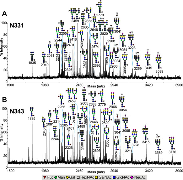
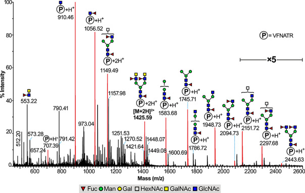
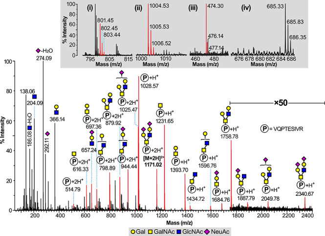
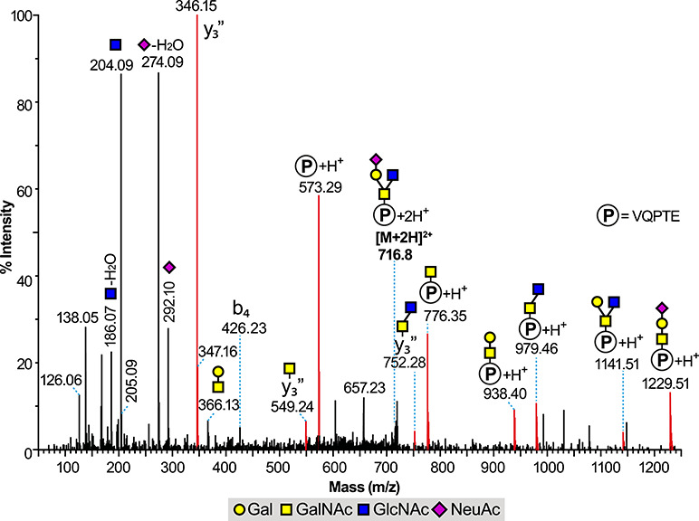
The novel coronavirus SARS-CoV-2, the infective agent causing COVID-19, is having a global impact both in terms of human disease as well as socially and economically. Its heavily glycosylated spike glycoprotein is fundamental for the infection process, via its receptor-binding domains interaction with the glycoprotein angiotensin-converting enzyme 2 on human cell surfaces. We therefore utilized an integrated glycomic and glycoproteomic analytical strategy to characterize both N- and O- glycan site-specific glycosylation within the receptor-binding domain. We demonstrate the presence of complex-type N-glycans with unusual fucosylated LacdiNAc at both sites N331 and N343 and a single site of O-glycosylation on T323.

Keywords: SARS-CoV-2; glycoproteomics; mass spectrometry; spike glycoprotein.


Figures

Fig. 1
Fig. 1
Glycomic analysis of the N331 and N343 glycopeptides derived from the RBD of the S1 spike SARS-CoV-2 glycoprotein. MALDI-TOF mass spectra of permethylated N-glycans derived from (A) 329-FPNITNLCPFGE-340 glycopeptide (containing the N331 glycosylation site) and (B) 341-VFNATR-346 glycopeptide (containing the N343 glycosylation site). All signals observed are singly charged sodiated ions [M + Na]+ and their structural assignment is based on monosaccharide composition, MS/MS fragmentation analyses and knowledge of the biosynthetic pathways. Residues shown on top of a bracket have not had their antenna location unequivocally defined.
Fig. 2
Fig. 2
Glycoproteomics analysis of the 341-VFNATR-346 glycopeptide derived from the RBD of the S1 spike SARS-CoV-2 glycoprotein containing the glycosylation site N343 found centered at 12.1 min in the LC elution profile. LC ES-MS (MSe mode) containing the [M + 2H]2+ molecular ion found at m/z 1425.59 and corresponding to the 341-VFNATR-346 glycopeptide with a core-fucosylated bi-antennary N-glycan with fucosylated LacdiNAc antennae (permethylated N-glycan found at m/z 2674 on Figure 1B and Supplementary Table SI). The peaks in red correspond to glycopeptide fragment ions deriving from the [M + 2H]2+ molecular ion. “P” inside a circle corresponds to the peptide backbone of 341-VFNATR-346. Area corresponding to m/z 2120 to 2600 has a 5-fold magnification. The residues shown on top of a bracket have not had their antenna location unequivocally defined.
Fig. 3
Fig. 3
Glycoproteomics analysis of the 320-VQPTESIVR-328 glycopeptide containing the putative glycosylation site at T325 and S325 derived from the RBD of the S1 spike SARS-CoV-2 glycoprotein. LC ES-MS (MSe mode) data containing the [M + 2H]2+ molecular ion found at m/z 1171.02 and interpreted as a single Core 2 substitution at Thr 323 with the HexNAc2Hex2NeuAc2 structure shown. “P” inside a circle corresponds to the peptide backbone of 320-VQPTESIVR-328. The insets correspond to zoom scan from the regions of (i) m/z 801, (ii) m/z 1004, (iii) m/z 474 and (iv) m/z 677.
Fig. 4
Fig. 4
Glycoproteomics analysis of the 320-VQPTE-324 glycopeptide derived from the RBD of the S1 spike SARS-CoV-2 glycoprotein. LC ES-MS/MS data (CAD MS/MS mode) of a doubly charged ion species (716.82+), showing clear evidence of T323 substitution with a mono-sialyl version (NeuAcHexHexNAc2) of the assigned Core 2 structure seen in Figure 3 and described in the text. Proline cleavage y” ions at m/z 346, 549 (y3-HexNAc) and 752 (y3-HexNAc-HexNAc) support the interpretation made. The unlabeled higher mass signals were devoid of 13C isotope clusters and not assigned as of glycopeptide origin.

References

    1. Ceroni A, Maass K, Geyer H, Geyer R, Dell A, Haslam SM. 2008. GlycoWorkbench: A tool for the computer-assisted annotation of mass spectra of glycans. J Proteome Res. 7:1650–1659. - PubMed
    1. Ciucanu I, Kerek F. 1984. A simple and rapid method for the permethylation of carbohydrates. Carbohydr Res. 131:209–217.
    1. Dell A, Morris HR. 2001. Glycoprotein structure determination by mass spectrometry. Science. 291:2351–2356. - PubMed
    1. Dewal MB, DiChiara AS, Antonopoulos A, Taylor RJ, Harmon CJ, Haslam SM, Dell A, Shoulders MD. 2015. XBP1s links the unfolded protein response to the molecular architecture of mature N-Glycans. Chem Biol. 22:1301–1312. - PMC - PubMed
    1. Do KY, Do SI, Cummings RD. 1997. Differential expression of LacdiNAc sequences (GalNAc beta 1-4GlcNAc-R) in glycoproteins synthesized by Chinese hamster ovary and human 293 cells. Glycobiology. 7:183–194. - PubMed

Publication types

MeSH terms